Juniper Publishers Journal of Case Studies is an international, peer reviewed open access journal, focussed on bringing out premium quality case reports in all areas of basic medical sciences and clinical specialities. ‘JOJCS’ aims to serve as a repository of medical case reports that can go a long way in enriching the professional knowledge of medical practitioners around the world.
Showing posts with label Peer Review journal of case reports. Show all posts
Showing posts with label Peer Review journal of case reports. Show all posts
It is well established that the etiology of allergic diseases is that combinations of mast cells and allergen-specific antibodies cause allergic symptoms when the patients meet allergens. Similarly, the etiology of auto-immune diseases is that combinations of cytolytic T lymphocytes and organ-specific antibodies cause injury of the organ. A most plain idea would be that break down of the above mentioned combinations must bring about disappearance of causes of the diseases. It seems to me that few, if any, contemporary Immunologists have such concepts. To work out the above mentioned concept, it is necessary to have the patients make non-specific antibodies by themselves. In order for the patients to do so, they need to receive intradermal injections with non-specific antigen preparations. Consequently, non-specific antibodies accumulate in the patients’ bodies, which may replace specific antibodies from respective cells bringing about elimination of causes of the diseases. Needless to mention, where there is no cause, there is no disease. Details are demonstrated elsewhere [1]. A possible alternative opinion may be transfusion with gamma-globulin solution. However, it is dangerous because anti-gamma-globulin antibodies may be produced in the recipient’s body, which could cause an anaphylactic reaction after a large number of infusions.
A 64-year-old woman (M.O.) visited my clinic on April 21, 2011. She told me that she had had a systemic urticaria with conjunctivitis and rhinitis in middle of May, 1987. She was diagnosed as systemic lupus erythematosus by a doctor in The Osaka Medical School Hospital after examination of her bone marrow in June, 1987. She received 40mg daily of oral prednisolon for 3 months, 10mg daily for 1 month, and 5mg daily continuously. I intradermally injected her at her upper-arms with 0.1ml of 1,000,000 fold with saline diluted Neurotropin; a product of Nippon Zohki Pharmaceutical Company (Osaka), consisting of an extract of rabbit skin inflamed by inoculation of Vaccinia virus, at 2~19 day intervals during the period from April 21, 2011 until August 25, 2011. The total number of injections was 18. She is enjoying a healthy life as of September 16, 2017.
Aim:Tumor grading is a significant predictor for clinical outcome of astrocytomas. The aim of the present study is the application of MIB-1 labeling index (LI) in differentiating Grade II and Grade III astrocytomas.
Methods:Our study included 50 cases of astrocytomas. MIB-1 LI was done in all cases and was compared in correlation with 2007 World Health Organization classification.
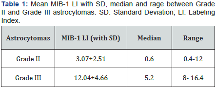
Result:The mean MIB-1 LI for Grade II astrocytomas was 3.07±2.51 and 12.04±4.66 in Grade III astrocytomas. P values were significant between Grade II and Grade III astrocytomas.
Conclusion:MIB-1LI is used as an effective tool in assessing astrocytic tumor proliferation and also as an important prognostic indicator supplementing the standard histopathological assessment. Present study demonstrates the usefulness of MIB-1LI in Grade II and Grade III astrocytic tumors where histopathologic grading becomes difficult based solely on morphology.
Astrocytomas constitute majority of primary intra cerebral tumors [1]. Tumor grading is a useful prognostic predictor in astrocytomas. The low- grade tumors are associated with better prognosis in comparison to high grade tumors. However, when small stereotactic guided biopsies are obtained, differentiation based only on morphology may be difficult and confusing. Therefore, MIB-1 Labeling index (LI) which determines the proliferation in astrocytomas have an important role to measure the diagnostic and prognostic clinical outcome [2].
MIB-1 antibody is an IgG1 class monoclonal antibody which recognizes a core antigen present in the nuclei of the cells in all phases of the cell cycle except in the resting phase [3]. World Health Organization (WHO) however does not include MIB-1 LI as a component in grading scheme for gliom as [4]. The cellular proliferation is related to tumor growth rate, quantification of this process has prognostic value and is related to tumor grading. The LI of any of the proliferating antigens is the number antigen positive cells divided by total number of cells sampled microscopic areas of the tumor. Ki67 are nuclear antigens that appear during the proliferative phases of the cell cycle [5]. Therefore proliferation rate can be assessed by measuring Ki67 labeling index using MIB-1 antibody. Hence MIB-1 is used in differentiation between diffuse and anaplastic astrocytomas.
This study signifies the importance of MIB-1LI in the assessment of Grade II and Grade III astrocytomas in correlation with histopathologic grading of astrocytomas based on WHO classification.
This study includes 50 patients diagnosed with Grade II and Grade III astrocytic tumors based on WHO classification of central nervous system (CNS) tumors over a period of four years from January 2007 to December 2010.
The specimens were processed by routine paraffin embedding technique. All the specimens were from biopsy of operated tumors. Paraffin sections of 3-5μ were obtained from each block and stained with hematoxylin and eosin (H and E). The tumors were graded according to the WHO classification of CNS tumors.
Our study included 29 patients of Grade II astrocytomas and 21 patients of Grade III astrocytomas which were analysed for MIB-1 LI by polymer labeling two step method using super sensitive polymer-Horseradish peroxidase (HRP).
MIB-1 (Ki-67 specific monoclonal antibody; BGX-297) Mouse monoclonal was used. Cases with small tissue size were not included in our study. Positive control used was Lymphoma. Immuno staining was evaluated at low power in tumor areas having the greatest number of immune reactive cells. Only nuclear staining was regarded positive.
The MIB-1 LI was indicated as the percentage of tumor cell nuclei stained, in areas of maximum staining and calculated in at least 1000 cells using a high power (40x) objective.
Survival data for length of survival was obtained for 33 patients over a period of 35 to 55 months and was evaluated based on the information obtained from clinical records on subsequent follow up. In the present study, a p value of <0.05 was considered significant for the performed tests. The ethical clearance was obtained prior to the study.
In the present study, age group ranged from 0 to 60 years. Third decade constituted the majority of the cases (50.54%) with male preponderance and male: female ratio of 1.4:1.
The majority of astrocytomas occurred in the middle cranial fossa (52%). Grade II astrocytomas constituted of 29 cases and mainly consisted of fibrillary type. On histopathology they were composed of fibrillary neoplastic astrocytes loosely woven together with mild degree of nuclear pleomorphism. Mitotic activity, necrosis and microvascular proliferation were absent.
Grade III astrocytomas or anaplastic type constituted of 21 cases and were characterized by increased cellularity, moderate nuclear pleomorphism, presence of mitotic figures, significant vascular proliferation and microcystic changes.
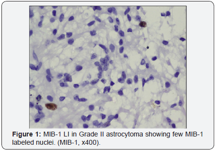
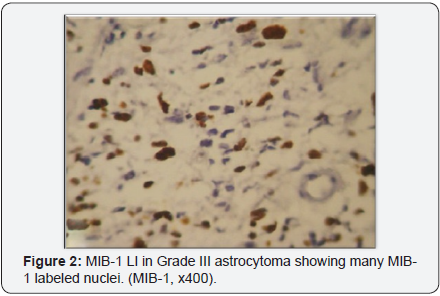
MIB-1 LI was significantly increased with the grade of astrocytoma. The mean MIB-1LI with a standard deviation along with median and range in Grade II and Grade III astrocytomas were analysed (Table 1) (Figure 1 & 2).
P values were obtained using Students unpaired T test. P value of <0.001 was obtained between Grade II and Grade III astrocytomas which were statistically significant. Survival analysis was done, but no significant confines were identified for MIB-1 LI. Better survival rate was observed in younger individuals, but was not significant based on statistical outcome.
The WHO classification of astrocytomas has limitations in predicting the clinical outcome and survival of patients. Literature studies have shown that proliferative index derived from MIB-1 LI provides more knowledge on the biological behaviour in various grades of astrocytomas directly impacting patient management and survival [6,7].
The result obtained in our present study, showed that third decade was the most common age group in the study population. The male preponderance with respect to astrocytomas is universally recognized, so as in our study [8]. Astrocytomas were usually supratentorial in adults and infratentorial in infants and children as observed by Pant et al. [5].
In the present study, Grade II astrocytomas where mostly fibrillary pattern and only two cases with gemistocytic type was identified. This finding was similar to those seen in studies by Pal et al. [9] and Wahal et al. [10].
Histopathologic diagnosis based on WHO classification of astrocytomas has been challenging to distinctly differentiate tumor grades either due to limited tumor material or due to difficulty in identifying mitoses in H and E stained sections [4].
In our present study, MIB-1 LI was done to assess the proliferative activity in Grade II and Grade III astrocytomas as it is sometimes difficult based on morphology and mitosis to differentiate between these two grades [11].
Johannessen & Torp [12] analysed and reviewed 16 studies which constituted a total of 915 patients demonstrating a substantial increase in MIB-1 LI with increasing grade of malignancy. As in our study, the reports demonstrated a mean of MIB-1 LI in Grade II and Grade III astrocytoma of approximately 3 and 12.
The statistical significance obtained in our study between Grade II and Grade III astrocytomas have been documented in various studies [2,13,14]. Rodrvguez et al. [15] and Hsu et al. [16] identified that Grade II tumors had significantly lower MIB- 1 LI compared to Grade III tumors. Arvinder et al. [17] study on gemistocytic astrocytomas found lower MIB-1 LI though they are classified as Grade II astrocytomas.
In a bivariant analysis done by Rodriguez- Pereira et al. [15] 19MIB-1 LI increased with age, histologic grade and a supratentorial location. Hence, the importance of the proliferation marker is influenced by many factors which reduce its value as an isolated prognostic parameter. Our study shows that MIB-1LI is dependent solely on the histologic grade and is independent of age and sex.
Most studies demonstrated positive correlations between MIB-1 LI, survival and recurrence. Paul et al. [18] found that gender, tumor location and radiotherapy had no significant association with survival. However, high MIB-1 predicted shorter survival and also an independent prognostic factor in Grade II astrocytomas.
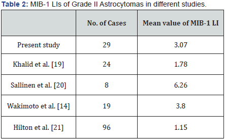
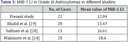
The mean MIB-1 LI in Grade II and Grade III astrocytomas in comparison with different studies are shown in (Table 2 & 3) [14,19-21].
A wide variety of factors were responsible for the differences in MIB-1 LI. These factors include fixatives used, technical issues with variety of detection procedures, dilutions of antibodies used, antigen retrieval procedures and finally interpretation of immune staining [14].
Grzy bicki et al. [22] conducted a inter observer variabilty study to assess the differences in MIB-1 LI and found that high levels limited the prognostic usefulness for patients with primary brain tumors. These key factors are important to be noted while planning the treatment options after considering MIB-1 LI in astrocytomas. MIB- 1 is an important clinical marker in astrocytomas, but with these shortcomings it is not reliable to use absolute cut-off values between different laboratories and it is also difficult to standardize it for prognostic purposes.
MIB-1 immuno staining worked well and yielded credible results in our study. Therefore, this marker when used in combination with clinical evaluation and tumor grade, provide a greater perspective in evaluating and strategic treatment planning with patients of astrocytomas.