Showing posts with label Transplantation Medicine. Show all posts

Monday, September 20, 2021

A Case Report of Widespread Melanoma with a Complete Response to Pembrolizumab- Juniper Publishers

 Juniper Publishers- Open Access Journal of Case Studies


A Case Report of Widespread Melanoma with a Complete Response to Pembrolizumab

Authored by Natalja Eigeliene

Abstract

Melanoma is an aggressive disease that accounts for approximately 2/3 of skin cancer-related deaths. In the past, treatment options for patients with advanced melanoma have been limited, with a poor response rate and failure to improve overall survival (OS). Immunotherapy is a promising treatment that has been shown to improve OS and, in many cases, produce durable remission in patients with advanced melanoma. Here, we report a case of a patient with widespread malignant melanoma at progression after biochemotherapy who achieved a durable complete response to pembrolizumab immunotherapy without major side effects. The patient has been progression-free and treatment-free for four years. This case adds to the evidence in support of early referral and initiation of pembrolizumab for patients who can tolerate it, with the intent to produce a sustained treatment-free response and preserved quality of life.

Keywords: Melanoma; Pembrolizumab; Immunotherapy; DOBS; Complete response

Abbreviations: DOBS: Lomustine, Bleomycin, Vincristine, Dacarbazine; I: Interferon; CT: Computer Tomography; MRI: Magnetic Resonance Imaging; FDG: Fluorodeoxyglucose; PET: Positron Emission Tomography; iv: Intravenous; sc: Subcutaneous

Introduction

Cutaneous melanoma, a type of skin cancer, has risen in incidence in recent years, according to the NCCN guidelines for cutaneous melanoma [1]. Unresectable or metastatic malignant melanoma has until recently been very difficult to treat. New therapies, including targeted therapies and immunotherapy, have been developed and implemented according to clinical guidelines [1]. Immunotherapy is an essential part of the treatment of metastatic and unresectable malignant melanoma [1]. Nevertheless, more research is needed to establish the optimal administration, treatment combination, and duration of therapy for different immunotherapeutic agents.

Case Report

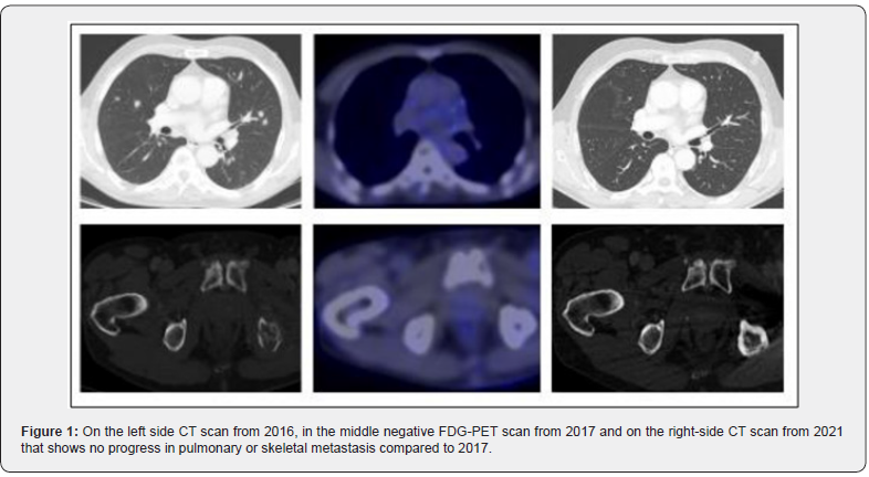
We present the case of a 70-year-old Caucasian man who reported with worsening pain in the lower back and left hip in 2016. He was an ex-smoker with no history of trauma to the back. The patient had psoriasis that had been in remission for several years. First, the patient sought treatment from a private doctor. Spine and SI-joint magnetic resonance imaging (MRI) was performed and revealed bone metastases. Thus, the patient underwent a body computer tomography (CT) scan, which showed multiple metastases in the thyroid gland, lungs, mediastinum, liver, and vertebrae Th4 and Th7 (Figure 1). A biopsy from the sacrum was obtained, and histology confirmed melanoma without BRAF mutation. The primary origin of the melanoma was not found.

Radiotherapy was given due to pain in the left hip (10 x 3Gy). Chemotherapy was then initiated with DOBS (lomustine, bleomycin, vincristine, dacarbazine)-interferon (I), which caused substantial side effects, such as weight loss, fatigue, nausea, and vomiting. Additionally, due to bone metastases, denosumab 120mg was given subcutaneously (sc). Three cycles of DOBS-I were given before treatment response was evaluated with a whole-body CT scan. Unfortunately, in the initial scan, the tumor was found to have progressed in the liver, mediastinum, and lungs. Thus, a decision to start second-line treatment with pembrolizumab was made.

Two hundred milligrams of pembrolizumab were given intravenously (iv) every third week. The patient had mild treatment-related side effects. After the second treatment with pembrolizumab, an itchy rash appeared on both lower extremities; a dermatologist diagnosed the rash as psoriasis. Local application of an anti-psoriatic cream comprised of calcipotriol and betamethasone was recommended and alleviated the rash. After the third pembrolizumab treatment in 2017, a whole-body CT scan revealed an excellent response to the treatment. Together, ten treatments were given triweekly, and FDG-PET-CT was performed (Figure 1). The patient achieved a complete response. Thus, pembrolizumab and denosumab were discontinued, and follow-up was initiated, including clinical examination, blood analyses, and regular CT scans. In spring 2020, the left hip joint was replaced by a prosthetic implant without any complications. The patient has attended regular follow-up visits at the dermatology and oncology clinics. To date, the patient has remained stable, with no signs of progression on the last CT scan on 2/2021 (Figure 1).
Discussion

Despite many years of research and some advances, metastatic malignant melanoma remains a significant challenge for modern medicine and is still regarded as incurable [2,3]. In our patient with widespread malignant melanoma, a durable (4 years) complete response to pembrolizumab immunotherapy as a second-line therapy for progression after radiotherapy and biochemotherapy was achieved. Follow-up is ongoing, without treatment, and no signs of cancer have been detected since 2017. There is a chance that this patient’s metastatic melanoma has been cured; however, it is too early to make that conclusion. These findings highlight the changes in active cancer treatments for solid tumors from life-extending treatments to curative therapies that can induce remission and potentially lead to cure, which requires life-long sustained remission [4].

A recent Cochrane analysis [5] demonstrated that immune checkpoint inhibitors improved progression-free survival rates compared to chemotherapy (HR 0.30, 95% CI 0.17 to 0.51) and that patients who received chemotherapy had worse PFS rates than those who received a combination of immune checkpoint inhibitors (HR 2.81, 95% CI 1.54 to 5.11) for melanoma.

The CR rate for chemotherapy is typically low, as shown in a meta-analysis including 68 chemotherapy trials for cancer in which the average CR rate was found to be 7.41% (5-10%) in solid tumors, e.g., colon, breast prostate, and melanoma (excluding testis cancer and pediatric leukemia) [6]. The CR rate has also been found to be low in patients with melanoma treated with single immunotherapeutics with a CR rate of 7.6% achieved with nivolumab [7] 1.7% with ipilimumab and 5-6.1% with pembrolizumab [7]. However, the CR rate improved up to 20% with pembrolizumab in a prolonged study in which five-year responses [8]. Currently, it is acceptable to discontinue checkpoint inhibitor treatment for patients who have achieved CR after six months of therapy, since the five-year risk of relapse is estimated to be less than 10% [8].

A durable complete response can be maintained after discontinuation of pembrolizumab in patients with metastatic melanoma. FDG PET/CT imaging can be used to assess tumor response to immunotherapy [9,10]. Our case report describes a clinical example of the successful use of PET CT imaging, as after the initial negative examination, the patient’s melanoma has not relapsed; however, the optimal timing and utilization of FDG PET/ CT imaging for decision making needs to be further investigated in prospective studies.

To know more about Juniper Publishers please click on: https://juniperpublishers.com/manuscript-guidelines.php

For more articles in  Open Access Journal of Case Studies please click on: https://juniperpublishers.com/jojcs/index.php


Wednesday, September 15, 2021

Effective Use of a Novel Biphasic Calcium Phosphate with Submicron Surface Topography in Posterolateral Spine Fusion - Juniper Publishers

 Juniper Publishers- Open Access Journal of Case Studies


Effective Use of a Novel Biphasic Calcium Phosphate with Submicron Surface Topography in Posterolateral Spine Fusion

Authored by Alwyn Jones

Abstract

The number of spine fusion procedures continues to increase with the aging population. Posterolateral spinal fusion (PLF) is one of the more challenging clinical indications in the spine requiring a bone graft because the aim is to form a large, consolidated bone mass through the paraspinous soft tissues with limited host bone contact. Synthetic calcium phosphate bone graft usage has increased in recent years, reducing the need to harvest large amounts of autologous bone. This class of bone graft closely resembles the composition of human cancellous bone and demonstrates a low incidence of adverse reactions and graft-related complications in the clinic. The body’s natural response to the tissue trauma associated with spinal surgery is the upregulation of macrophages of the pro-inflammatory M1 phenotype. This can lead to the formation of scar tissue and failed fusion. This biphasic calcium phosphate (BCP) bone graft with a unique submicron needle-shaped surface topography has been shown in in vitro studies of human-derived monocytes to promote attachment and spreading of anti-inflammatory M2 macrophages, leading to the formation of bone instead of scar tissue. This report on a consecutive case series assessed radiographic, functional and pain outcomes following posterolateral lumbar fusion using a novel biphasic calcium phosphate bone graft with a unique submicron topography in a potentially bone-density-challenged group of female patients.

Keywords: Spine fusion; Bone graft; Perimenopausal; Anteroposterior, Lateral radiographs; Biphasic calcium phosphate

Introduction

The number of spine fusion procedures continues to increase proportionately with the aging population [1,2]. Posterolateral spinal fusion (PLF) is one of the more challenging clinical indications in the spine requiring a bone graft because the aim is to form a large, consolidated bone mass through the paraspinous soft tissues with limited host bone contact. A major obstacle associated with this indication is the lack of available local autograft. As a result, surgeons continue to search for the most advanced and affordable bone graft to achieve successful spine fusion without the well-known co-morbidities related to harvesting autograft [3]. Synthetic calcium phosphate bone graft usage has increased in recent years as it has demonstrated support of bone formation and reduced the need to harvest large amounts of autologous bone [4,5]. This class of bone graft closely resembles the composition of human cancellous bone and has proven to be cost-effective with a very low incidence of adverse reactions and graft-related complications [6].

The body’s natural response to spinal surgery is the production of macrophages, especially the pro-inflammatory M1 phenotype [7,8]. This can lead to the formation of scar tissue and, ultimately, a failed fusion. MagnetOs is a biphasic calcium phosphate (BCP) bone graft with a unique submicron surface topography. Its submicron needle-like surface features have been shown, using in vitro studies of human-derived monocytes, to promote the attachment and spreading of M2 macrophages, leading reliably to the formation of bone instead of scar tissue [9].

In preclinical studies, the BCP has been shown to promote bone formation, even in soft tissue, without the need for added cells or growth factors [10]. This novel BCP is designed to mimic the porous, trabecular structure of cancellous bone. Clinically relevant animal models of posterolateral fusion demonstrate that bone formation take place throughout MagnetOs simultaneously, leading to uniform, solid and stable fusions [11,12].

The purpose of this evaluation of a consecutive case series was to assess radiographic success as well as functional and pain outcomes following posterolateral lumbar fusion using a novel biphasic calcium phosphate bone graft with a unique submicron topography (MagnetOs™, Kuros Biosciences, B.V.).

Four female patients, with an average age at the time of surgery of 62.7 (50-70 years of age), underwent a single or a two-level posterolateral lumbar fusion procedure for the treatment of degenerative spondylolisthesis or deformity using MagnetOs over the transverse processes and/or the facet joints. Each of these cases involved a perimenopausal/postmenopausal woman; a life stage known to negatively affect bone quality and cause concern when aiming for a successful fusion outcome.

Anteroposterior and lateral radiographs, visual analog scale (VAS) for back and leg pain, the Oswestry Disability Index (ODI), and medication usage were reviewed pre-operatively and post-operatively at 6 and 12 months. VAS and ODI scales were not available at 12 months; however, considering the marked improvement reported by each patient at 6 months, minimal, if any, significant improvement at this time point would be expected at 12 months.

Case Reports

Case 1: ALIF

A 69-year-old woman presented with back and left leg radicular pain. Conservative treatment included acupuncture, nerve blocks and physical therapy. Preoperative radiographs and MRI findings revealed lumbar scoliosis and foraminal stenosis at L5-S1. The patient-reported an ODI score of 56 and back pain scores equal to leg pain (8/10) on VAS. Pain was also managed with a combination of Ibuprofen and acetaminophen. Surgical treatment included an anterior interbody/posterior fusion with posterior fixation at L5-S1 and MagnetOs granules mixed with bone harvested from the facets placed laterally to the posterior stabilizing hardware.

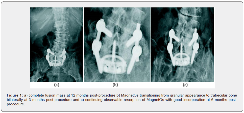
The patient's leg pain had completely resolved by the 6-month visit. The patient reported completely resolved leg pain, reduced back pain (4/10), an ODI function score of 34 and medication usage had been reduced significantly. Radiographs revealed fusion progression with good incorporation of BCP in the posterior fusion mass. Bone remodeling and graft resorption were evidenced by the loss of granular appearance graft. The 12-month-visit radiographs showed solid bilateral bridging fusion. The patient indicated all pain medication had been discontinued and clinically, no further surgery was indicated (Figure 1).

Case 2: PLIF

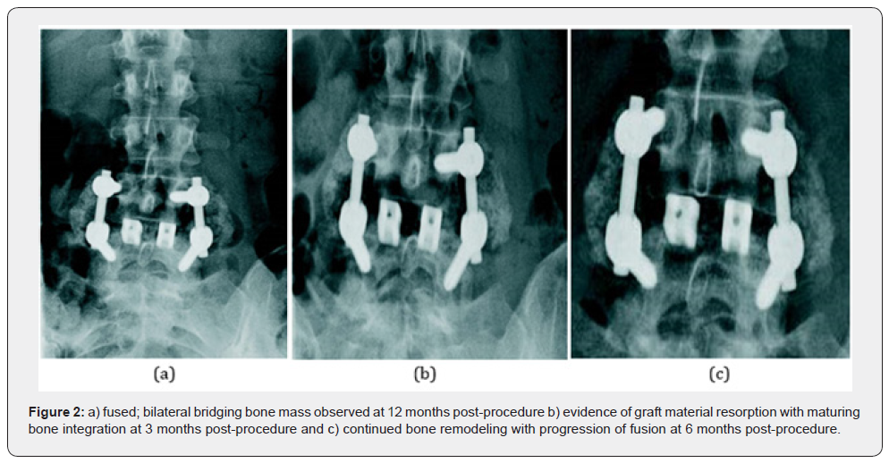
A 50-year-old female presented with intractable 10/10 back pain and 8/10 right leg pain on VAS, pre-operatively. Following failed conservative care, which included MS Contin and Tramadol for pain, nerve blocks and physical therapy, surgery was indicated. Preoperative radiographs and magnetic resonance imaging (MRI) revealed degenerative spondylolisthesis at L4-5. She underwent a posterolateral fusion with instrumentation at L4-5 with MagnetOs granules mixed used as an extender over the lateral gutters combined with a posterior lumbar interbody fusion at L4-5 using a PEEK coated interbody device packed with local bone. Imaging at 6 months showed the progression of fusion with good bone remodeling and the development of bilateral bridging. The patient reported minimal back pain (3/10) and leg pain (1/10) requiring only Tramadol at 50mg daily. Her function score dropped from 54 pre-operatively to 20. At the 12-month visit, radiographs demonstrated a solid bilateral fusion and Tramadol only as needed (Figure2).

Case 3: XLIF

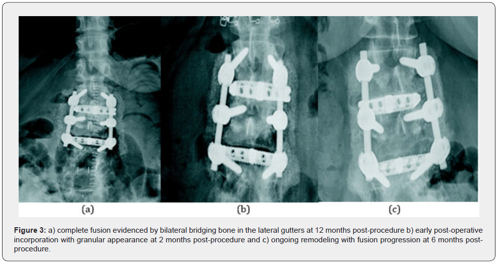
A 62-year-old female with back pain, bilateral leg pain, and paresthesia. She required the use of a wheelchair to ambulate and has resultant paraspinal muscle atrophy. She had lumbar fusion surgery at L4-5, L5-S1 10 years prior. The patient-reported pre-operative 10/10 back pain, 9/10 leg pain, and a very poor ODI function score of 78. Conservative care included physical therapy and pain medication through a combination of Duragesic, Duloxetine, and Amitriptyline. MRI and plain radiographs showed degeneration at L2-3, degenerative spondylolisthesis at L3-4, and a fusion L4-S1 with loss of lordosis. The surgical plan involved extending the fusion above the previous fusion levels and decompression at L3-4. A 2-level posterolateral fusion with fixation at L2-3, L3-4 placing MagnetOs granules, which were mixed with local bone, in the inter-transverse process area was performed in combination with interbody fusion at the same levels. Radiographs at 6 months demonstrated fusion progression, good incorporation of the BCP into the fusion bed and graft resorption evidenced by the change from granular to more trabecular appearance. The patient was still rehabilitating but reported significantly reduced pain and improved function scores. Her back pain score decreased from 10/10 to 4/10 on the VAS, leg pain decreased from 9/10 to 1/10, and her ODI score improved from 78 to 40. Pain medication usage reflected the reported improvement in pain, as narcotics had been discontinued. At 12 months, the radiographs revealed a solid fusion bilaterally and medication had been reduced to Tramadol 50mg as needed (Figure 3).

Case 4: TLIF

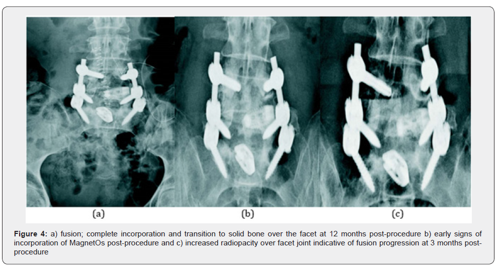
A 70-year-old female with a history of prior decompression at L4-5 and fusion at L5-S1 presented with back pain greater than leg pain. She had not benefited from conservative treatment consisting of physical therapy and pain management. The patient-reported 8/10 back pain, 2/10 leg pain, and a poor ODI function score of 60. Preoperative radiology reports revealed degenerative spondylolisthesis at L4-5. She underwent a posterolateral fusion with fixation at L4-5. BCP granules were mixed with bone harvested from the facets and placed medially to the posterior stabilizing hardware. The surgical plan also included an interbody fusion at L4-5. Radiographic imaging at three months post-op demonstrated fusion progressing well, good graft incorporation and resorption with noticeable changes from granular to a more trabecular appearance. The patient-reported a marked reduction in pain and improved function scores. Her back pain had decreased from 8/10 to 1/10 on the VAS scale, leg pain was completely resolved, and her ODI function score improved by a reduction from 60 to 20. All pain medications had been discontinued other than OTC Tylenol, as needed. At 12 months, the x-rays demonstrated solid fusion and full incorporation of bone graft as demonstrated by increased radiopacity at the facets (Figure 4).

Discussion

Historically, conventional synthetic bone grafts such as β-TCP have underperformed when compared to autograft in spinal fusion [13]. The cases described herein clearly demonstrate the effective use of biphasic calcium phosphate with a novel submicron surface topography in achieving fusion in the posterolateral spine. This cohort includes four (4) women of menopausal/post-menopausal age at risk for diminished host bone quality. BCP granules were implanted over the facets and/or in the lateral gutters. It is the posterolateral fusion procedure that presents an additional challenge for fusion, as in this defect, the bone must form over the farthest distance from the host bone.

Each case showed successful outcomes radiographically and in pain and function scores. Regular follow-up x-rays showed the replacement of the granular nature of the graft with trabecular bone over time. Significantly reduced pain and function improvements were demonstrated with a 67.5% average (range 50%-90%) reduction in back pain and a 95% average (range 90-100%) reduction in leg pain. The functional scores improved by an average of 31.5 points (range 22-40 points) and pain medication usage was dramatically reduced or eliminated. No device-related adverse events were reported and none of the patients required revision surgery within 12 months following the index procedure.Despite this being a small cohort of patients, these preliminary findings indicate that this BCP is an effective bone graft for posterolateral fusion, worthy of future clinical research.

This case series review demonstrates successful fusion results in a potentially bone-density-challenged group. It shows that MagnetOs directs bone formation early in the healing process and the graft resorbs at a physiologically balanced rate, thus supporting the transition from woven to trabecular bone and the development of uniform, stable and solid posterolateral fusions. These outcomes confirm results from preclinical studies that the BCP is an ideal bone graft extender for use in posterolateral fusion.

To know more about Juniper Publishers please click on: https://juniperpublishers.com/manuscript-guidelines.php

For more articles in  Open Access Journal of Case Studies please click on: https://juniperpublishers.com/jojcs/index.php


Juniper Publishers: A Leading Open Access Platform for Global Research in 2025

  Juniper Publishers: A Leading Open Access Platform for Global Research in 2025   Juniper Publishers: A Leading Open Access Platform fo...