Tuesday, July 30, 2019
Juniper Publishers: Internet Computerized Adaptive Testing to Detect C...
Juniper Publishers: Internet Computerized Adaptive Testing to Detect C...: Psychologyand Behavioral Science International Journal Juniper Publishers Abstract Objective: Surveys are often conducted but rar...

Friday, July 26, 2019
Facial Nerve Palsy after General Anaesthesia, A Less Reported Complication: Case Report-Juniper Publishers
Juniper Publishers-Open Access Journal of Case Studies

Facial Nerve Palsy after General Anaesthesia, A Less Reported Complication: Case Report
Authored by Rajesh Chaudhary
Abstract
Peri-operative nerve injuries are less documented but known complications. They are more common after regional anaesthesia but may be seen after general anaesthesia too. Peripheral branches of facial nerve are likely to be injured due to pressure exerted during bag and mask ventilation or due to stretch exerted while pulling the jaw forward. Most of the times these injuries are transient and recover within the time period of few days to weeks without any residual effects or they could be serious and permanent. Difficult intubation may invite trouble and cause facial nerve injury.
Keywords: Facial nerve palsy; Neuroprexia; Peripheral nerve injuries; Regional anaest
Introduction
Injury to the peripheral nerves in the peri-operative period is a rare but sometimes serious, documented complication. These injuries are a source of great anxiety and morbidity to the patients and their families besides they are leading cause of insurance claims. According to one data these injuries are a cause of litigation against anaesthetists as high as 16% [1]. These injuries may be minor and transient or permanent. Although there exact incidence is not known but one study has put the permanent nerve damage after surgery and anaesthesia to be somewhere between 0.03%-1.4%. Facial nerve injury may be seen after general anaesthesia which usually improves with the passage of time leaving the permanent damage only in a minor number of patients. The nerve is vulnerable to trauma due to variability in its course or sometimes because of its superficial nature, which makes it vulnerable to pressure and stretch [2]. Use of inappropriate size laryngeal mask airway(LMA) or endotracheal tube or a difficult intubation increase the chances of injury as highlighted by this case .
Case
We present the case of a 48 year old male weighing 65 kg who was posted for elective cholecystectomy. There was no history of any co morbidities but a difficult intubation was anticipated by the anaesthetist in view of short neck and a large tongue. At the operating table the induction of anaesthesia was started by the anaesthetist with 2 (mg) miligram/kg of Propofol along with 2 microgram/kg of Fentanyl and 0.5 mg/kg of Succinylcholine was given intravenously after preoxygenation with 100% oxygen for three minutes with bag and mask. An Endotracheal tube of size 7.5 was used but the trachea could not be intubated.
A size 6.5 tube also could not be negotiated into the trachea of the patient. Meanwhile the patient was again ventilated with 100% oxygen and laryngeal mask airway of size 4 was tried after proper lubrication with lignocaine jelly by another anaesthetist but the procedure again failed even after two attempts. The procedure was abandoned and patient shifted back towards after recovery. About six hours later the patient was seen in the wards. Patient complained of perioral numbness and difficulty in speaking. Angle of mouth was deviated towards left side. No other neurological sign was seen.
Anticipating some cerebrovascular accident plain CT scan of the head was done and it was found to be normal. The case was discussed with the physician also but no obvious cause could be found. We had never seen such a condition before hence extensive search of literature available over internet was made. A transient injury of the facial nerve was kept as the diagnosis. Prognosis was explained to the patient and counselling done regarding future outcome of the condition. Within a week the patient made full recovery and is still in follow up.
Discussion
The perioperative peripheral nerve injuries are more common after regional anaesthesia techniques but are seen after general anaesthesia too. Different nerves injured in decreasing order of frequency are ulnar nerve, brachial plexus, lumbosacral root, spinal cord, sciatic nerve, median nerve, radial nerve and femoral nerve respectively. Facial nerve injury is further rare [3]. Peripheral facial nerve injury is a mononeuropathy which is more commonly idiopathic or primary as compared to secondary to known causes like trauma, tumors, ischemia, viral infections or local anaesthetic agent toxicity [4]. Facial nerve is primarily a motor nerve with small sensory component. Facial nerve emerges out of the skull through stylomastoid foramen and becomes superficial to the mandible by entering the parotid gland where it gives off temporal, zygomatic, buccal, marginal mandibular and cervical branches. The main trunk may give off branches at different level in relation to the parotid gland. Digital pressure behind the mandible or excessive pressure exerted by the facemask can cause a traumatic lesion of the facial nerve. Sometimes the branches of facial nerve may be superficial to the parotid gland making the buccal and marginal mandibular branches especially vulnerable to injury during bag and mask ventilation for general anaesthesia [5].
On the basis of nerve pathology, Seddon [6] classified the nerve injuries into three categories. Neurapraxia is the simplest of injuries with damage only to the myelin sheath and an intact axon. The chances of recovery are excellent. When the axon is damaged but the endoneurium and supporting connective tissue framework is preserved, it is called Axonotmesis. The chances of recovery are variable. Neurotmesis is the most severe injury where the nerve is completely destroyed along with the connective tissue network and the chances of recovery are almost negligible [6].
Facial nerve injury could be because of unknown (Idiopathic) or known (Secondary) causes. Idiopathic facial nerve injury is more common [4]. Various mechanisms of nerve injury are ischemia, direct trauma, stretch and compression, and drug toxicity. Patients having diabetes mellitus, smokers, poor general health, pre-existing neuropathies are more prone to nerve injuries. These injuries are more common in neurosurgery, cardiac surgeries, and orthopaedic surgeries and with longer hospital stays. Endotracheal intubation and laryngeal mask airway (LMA) are known to cause nerve injuries.
Trauma while insertion or using a small size tube or LMA should be avoided. Inappropriate cuff size increases the chances of injury by exerting more pressure to occlude the airway [2]. Sometimes the jelly used while insertion of LMA may cause transient nerve paralysis. Onset of symptoms may be immediate in case of direct trauma to the nerve or may be delayed in case of inflammation and drug toxicities. The diagnosis is mainly clinical aided by MRI (Magnetic resonance imaging) and nerve conduction studies. Generally the recovery of peripheral facial nerve injuries is good with a complete recovery in about 85% over a period of weeks to months while as much as 15% may suffer a permanent damage leading to weakness, synkinesis, facial spasms, contractures, decreased tears or psychosocial effects [4].
Conclusion
The peripheral nerve injuries in the perioperative period are a less documented complication .They are a source of great discomfort for the patients and their families as well as a matter of litigation against the doctors. They are very rarely seen after general anaesthesia as compared to regional anaesthesia. Their incidence may be under reported and less but every reported case will add to the better understanding, early and good management of these injuries. They may be very less in context of total perioperative complications but are lifelong for those who suffer permanent damages. One should be aware about the possibility of a nerve injury. Patients should be counselled about the injury and it’s possible outcomes and followed up from time to time. A good sense of observation and awareness of a possible nerve injury may prevent such complications.
For
more Open Access Journals in Juniper Publishers please
click on: https://juniperpublishers.com/journals.php
For
more articles in Open Access
Journal of Case Studies please click on: https://juniperpublishers.com/jojcs/
To
know more about Peer
Review Journal of Case Studies
Wednesday, July 24, 2019
Epidemiology of Adenoid Cystic Carcinoma of the Breast in a Developing Community-Juniper Publishers
Juniper
Publishers-Open Access Journal of Case Studies

Epidemiology of Adenoid Cystic Carcinoma of the Breast in a Developing Community
Authored by Wilson IB Onuigbo
Abstract
A well recognized rare breast entity is the adenoid cystic carcinoma [ACC]. Indeed, a few series have long been reported from several parts of the world. Now, according to the Birmingham [UK] group, there is the concept that the histopathology data pool promotes epidemiological analysis. Therefore, the purpose of this paper is to present from a developing community the data collected from such a pool with reference to women of the Ethnic Group called the Ibos or Igbos, who live mainly in the South-eastern region of Nigeria. Of the sixteen collected examples, most were gathered from the Cosmopolitan City of Enugu. Both sides were affected equally. The 31-50 age groups preponderated, while a particular surgeon dominated the operation scenery. Distant practitioners profit by sending biopsies to a centrally located Reference Laboratory.
Keywords: Breast; Type; Carcinoma; Adenoid cystic; Distance; Ibos; Enugu; Niger
Introduction
During the 1970s, there was a rise in the reprint request [RR] traffic. Thus, I freely received during that time single or few case reports as reprints on ACC at my request [1-5]. Incidentally, the microphotographs were compelling, although there were some variations. By 1980, Macartney, Rollaston and Codling [6] postulated from Birmingham (UK) that the establishment of a histopathology data pool would facilitate epidemiological analysis. Fortunately, this idea was in consonance with my own work in those days.
Investigation
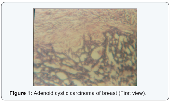
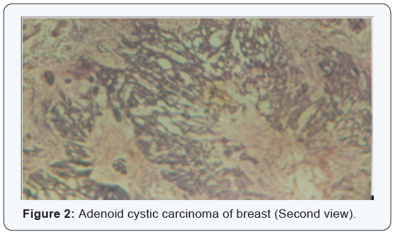
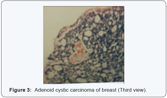
During the 1980s and 1990s, the author was privileged to head a Reference Pathology Laboratory situated in Enugu, the capital city of the then Eastern Region of Nigeria. Its histopathology data pool yielded 506 breast carcinomas, 16 being diagnosed personally as ACC. They are analyzed hereunder. Illustrative photographs are also adjoined (Figure 1-3).
Results
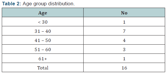
Tables 1 and 2 show the epidemiological analyses. It is evident
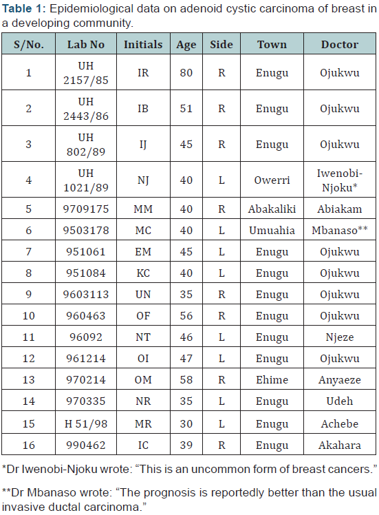
- that the lesions featured equally on both sides,
- most specimens were from the cosmopolitan city of Enugu,
- there was preponderance in the 31-50 age groups and
- Surgeon Ojukwu took pride of place.
Discussion
Among the RRs of the 1970s, let me select, on purpose, the careful work of Anthony and James [5]. They presented 3 cases from 2686 breast cancers within a period of 10 years. Moreover, as they agreed, “The term adenoid cystic carcinoma is the one recommended by the World Health Organization in preference to other synonyms.” Their range of photographs confirmed the requisite appearances. Also noteworthy was its tendency to carry a uniquely favorable prognosis when compared with similar tumors elsewhere in the body.
In the local context, ACC was diagnosed in a 68-year-old surgeon well over a decade ago as regards his bowel cancer; it has till now proved truly prognostic [7]. Also, I have found it to be a molecular variant in local albino skin cancers [8]; their own follow up should also be fruitful.
Concerning the recent world literature, what are some pointers? From India, a 40-year-old female with final histopathologic diagnosis of ACC underwent adjuvant external beam radiotherapy and was on follow-up without recurrence for more than a year [9]. From Seoul, Korea [10], the report on 6 patients was to the effect that “Although some of our patients developed local recurrence or distant metastases, all patients had favorable clinical course, and to date, none of the patients has died from complications of her disease.” Concerning a joint Korea-USA study [11], their comprehensive review provided experience with the ACC of the breast as well as an overview of clinical, histopathological, and molecular genetics, features. From the Mayo Clinic in USA [12], the conclusion was clear, namely, “Recognition of ACC is important to avoid delay in diagnosis because this tumor has a good prognosis with rare metastases to axillary lymph nodes. Axillary nodal sampling by fine-needle aspiration or core biopsy is rarely indicated.”
A population-based cohort study of ACC in the United States (1977 to 2006) was conclusive [13]: Breast-ACC among women is characterized by ER-negative/PR-negative expression, rare regional lymph node involvement, a favorable prognosis with excellent survival, and absence of associated cancers. These findings reinforce the importance of tailored treatments for breast-ACC and lend credence to the apparent heterogeneity of basal-like breast cancers. Incidentally, there was debate in the UK as regards the benefit accruable to practitioners distant from a Reference Laboratory [14]. Indeed, the experience in this developing community is that of its desirability [15].
For
more Open Access Journals in Juniper Publishers please
click on: https://juniperpublishers.com/journals.php
For
more articles in Open Access
Journal of Case Studies please click on: https://juniperpublishers.com/jojcs/
To
know more about Peer
Review Journal of Case Studies
Subscribe to:
Posts (Atom)