Juniper Publishers- Open Access Journal of Case Studies
Estimation of Superficial Venous Reflux with Duplex Ultrasound and Foot Volumetry
Authored by Helene Zachrisson
Abstract
Objective: To evaluate quantitative duplex ultrasound (DUS) parameters of reflux in patients with isolated great saphenous vein insufficiency.
Methods: 20 limbs were studied. DUS derived reflux time (RT, sec), peak reflux velocity (PRV, cm/s) and reflux volume flow (ml/min) were evaluated and related to expelled volume (EV, ml) and half refilling time (T50, sec) measured by water-based foot volumetry with and without compression of superficial veins.
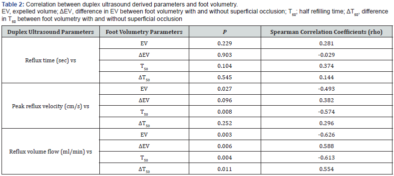
Results: Reflux volume flow correlated significantly to all hemodynamic parameters assessed by foot volumetry, i.e., EV (p = 0.003), ΔEV (p = 0.006), T50 (p = 0.004) and ΔT50 (p = 0.011). PRV displayed a weaker correlation to foot volumetry parameters EV (p = 0.027) and T50 (p = 0.008). No significant correlation was found between RT and foot volumetry.
Conclusion: These results indicate that reflux volume flow may be a potential parameter in future attempts to quantify reflux using DUS in patients with isolated great saphenous vein insufficiency.
Keywords: Venous insufficiency; Foot volumetry; Duplex ultrasound; Pathophysiology; Anatomical distribution
Introduction
Chronic venous insufficiency is a common condition with clinical signs ranging from minor telangiectasias, varicose veins, edema to more severe stages with skin manifestations as eczema, lipodermatosclerosis and venous ulcers [1-3]. The diagnosis relies on physical examination (C of the CEAP classification, “Clinical Etiology Anatomy Pathophysiology”) [4] as well as noninvasive testing [3,5]. Duplex ultrasound (DUS) is considered to be gold standard and provides diagnostic information about the anatomical distribution of the disease [3,6]. A retrograde flow (reflux time, RT) of more than 0.5 seconds is generally used to define the presence of reflux [6]. However, individual RT does not seem to reflect the magnitude of reflux and the correlation between severity of disease or hemodynamic state and RT is limited [7]. Based on this it has been suggested that RT may be used for detection of reflux but other DUS derived parameters are needed for quantifying venous insufficiency [7,8]. Previous attempts to quantify reflux using DUS has involved peak reflux velocity (m/s), calculated reflux volume flow (ml/min) and reflux volume (ml) [7], however, the optimal method for quantifying reflux by DUS is still unclear [9]. Quantitative information of global venous hemodynamics can be derived from plethysmographic measurements such as strain gauge, photo, air as well as foot volumetry [10-12]. We have shown that it is possible to predict post-interventional outcome in Great Saphenous Vein Incompetence using strain-gauge plethysmography [10].
Foot volumetry may provide accurate information on the magnitude of global venous reflux as well as correlate to C in the CEAP classification [4,12]. The aim of the study was to evaluate DUS derived reflux parameters in patients with isolated great saphenous vein insufficiency (GSV) compared to quantifying plethysmographic measurements using foot volumetry.
Material and Methods
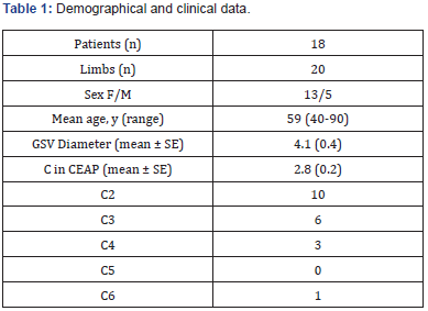
46 consecutive patients referred to Department of Clinical Physiology, Linköping University Hospital for evaluation of venous insufficiency in the lower limb were evaluated according to the study protocol. All patients were investigated with both Duplex ultrasound (DUS) and water-based foot volumetry. Six patients presented with small saphenous vein (SSV) insufficiency. Patients with mixed and/or isolated SSV insufficiency were excluded from the study. Finally, eighteen patients with isolated great saphenous vein (GSV) insufficiency (13 women and 5 men, mean age 59 years, range 40 – 90 years), two with bilateral GSV insufficiency (20 legs) were included in the study. Demographical and clinical data (C in CEAP) [4] is presented in Table 1. The study was approved by the regional ethical review board in Linköping, Sweden, and written informed consent was provided by each participant.

Duplex ultrasound
DUS examinations were performed with ACUSON S2000 system (Siemens Medical Solutions, Malvern, PA, USA) with 9 and 18MHz transducers. The 9MHz transducer was used for assessment of reflux. Patients were examined in the sitting position and superficial (saphenous veins and tributaries), perforator and deep veins (femoral, common femoral, deep femoral, popliteal, and calf veins) were scanned in both longitudinal and transverse planes. A distal manual compression was used to determine the valvular integrity. Normal veins were defined as veins with no reflux, normal reflux time (RT, duration < 0.5sec), or a very short reflux area (between 2 or 3 valves). If a pathologic reflux was detected, i.e., RT > 0.5sec, spectral Doppler measurements was performed along the vein in the longitudinal plane at 60o angle. Measurements were made in the insufficient GSV at the distal thigh level. At least three measurements were conducted at each point (proximal, mid and distal part on the thigh) and the mean value was used in the calculations. The anatomical extent of reflux was carefully noted, and no perforator reflux was noted on the calf or thigh level. Spectral flow velocity and vessel diameter was measured where the vein was straight, and turbulent areas were disregarded. The magnitude of reflux was quantified in several ways. i.e., RT (sec), peak reflux velocity (PRV, m/s) and time average velocity (TAV, m/s) during the first second of reflux. The vessel lumen was considered circular and the cross-sectional area was calculated as the area of a circle (A = Πr2 ) . Thus, reflux volume flow (ml/min) was calculated according to the following:
reflux volume flow (ml / min) = TAV (m / s) × A (cm2 ) × 60.
Hence, three DUS derived parameters were studied: RT (sec), PRV (m/s) and reflux volume flow (ml/min).
Foot volumetry
Global venous hemodynamics were evaluated with water- based foot volumetry [13]. The foot volumeter consisted of an open, water-filled box, which enabled measurements of volume changes during exercise [13]. Patients performed 20 knee bends at the rate of one every two seconds, and after the exercise phase was completed the patients remained completely still during the refilling phase. Expelled volume (EV, ml), and the time taken in seconds for 50% (T50) of the venous volume to be refilled was evaluated both with and without compression of superficial veins either above or below knee. A 10cm wide tourniquet was inflated to 60mmHg to achieve superficial compression.
Statistics
Values are expressed as mean ± SD unless otherwise stated. Spearman correlation coefficient (rho) were calculated for non-parametric linear association between different DUS parameters and foot volumetry measurements. p-values < 0.05 were considered significant. Statistical analyses were carried out using SPSS 24.0 for Windows (Armonk, NY: IBM Corp.).
Results
Isolated great saphenous vein (GSV) insufficiency was detected in 20 legs. DUS measurements displayed a mean vessel diameter of 4.2 ± 2.0mm. RT was 3.3 ± 1.3sec, PRV 45 ± 19cm/s, TAV 13.0 ± 6.9cm/s, and calculated reflux volume flow 128 ± 114ml/ min. EV was 13.6 ± 7.9ml, and T50 was 7.3 ± 6.4sec during foot volumetry without superficial compression. EV increased to 20.1 ± 8.1ml, and T50 increased to 13.0 ± 6.2sec after superficial occlusion. The calculated difference between foot volumetry measurements with and without superficial occlusion, i.e., ΔEV and ΔT50 were 6.5 ± 9.6ml and 5.8 ± 5.0sec respectively.
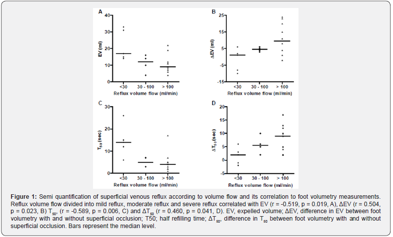
Table 2 shows the correlation between different DUS derived parameters and foot volumetry measurements. Reflux volume flow correlated negatively with foot volumetry parameters EV (rho = -0.626, p = 0.003) and T50 (rho = -0.613, p = 0.004). Reflux volume flow correlated positively with ΔEV (rho = 0.588, p = 0.006) and ΔT50 (rho = 0.554, p = 0.011). A negative correlation was also found between PRV and EV (rho = -0.493, p = 0.027) as well with T50 (rho = -0.574, p = 0.008). No correlation was detected between RT and foot volumetry parameters. Figure 1 shows reflux volume flow divided into three groups, mild refux (< 30ml/min), moderate reflux (30-100ml/min) and severe reflux (> 100ml/ min). This classification demonstrated a correlation with EV (rho = -0.519, p = 0.019), ΔEV (rho = 0.504, p = 0.023), T50 (rho = -0.589, p = 0.006) and ΔT50 (rho = 0.460, p = 0.041).


Discussion
This study was designed to evaluate different DUS parameters describing reflux in patients with isolated GSV and relate them to alterations in global venous hemodynamics based on water-based foot volumetry. The main findings were that particularly DUS derived reflux volume flow significantly related to global hemodynamic measurements during foot volumetry. Thus, the present study supports previous findings that reflux volume flow may be a potential parameter in future attempts to quantify reflux using DUS in patients with isolated GSV insufficiency.
DUS is a well-established method in the diagnosis of CVI and is able to measure several reflux components [6]. There has been numerous attempts to use one or more of these components as indexes for global reflux severity, although the most useful and reliable parameter for quantification of superficial reflux is still under debate. On the other hand, dynamic measurements of volume changes with plethysmographic methods may provide accurate quantitative information about whole limb venous haemodynamics and abnormalities in the reflux phase [10-12]. Foot volumetry was used in this study and previous findings suggest that it may provide accurate information on the magnitude of global venous reflux as well as correlate both to the clinical severity of the disease and the ambulatory venous pressure [12,14]. A further advantage with foot volumetry is that the method uses a dynamic and quite heavy situation, kneeling, which is supposed to simulate walking. In order to investigate the relation between DUS parameters and global reflux occlusion of the superficial system was accomplished with a tourniquet to evaluate the magnitude of superficial reflux in each patient.
RT was initially viewed as potential quantitative parameter and earlier studies found that patients with ulceration demonstrated a longer total mean RT than those without ulcers [15]. In the present study, no association was found between RT and any parameter of global reflux derived from foot volumetry. This is consistent with more recent studies who have shown RT to be a poor quantifier of individual reflux as well as an inadequate parameter to discriminate between disease severity [7,8,16]. Taken together, RT may primary be viewed as a qualitative parameter. Other parameters, such as, PRV has been found to provide a better quantitative evaluation compared to RT, e.g., PRV > 30cm/s has in previous studies been suggested as a risk factor for venous ulceration and patients with skin changes presented with significant higher PRV [17,18]. In line with this we found that PRV was associated to foot volumetric parameters EV as well as T50.
The parameters derived from DUS that showed best correlation with foot volumetry measurements was volume flow. Reflux volume flow was related to EV and T50 at baseline as well as to changes in global reflux after occlusion of the superficial veins evaluated by ΔEV and ΔT50. Assessment of retrograde flow volume has previously shown that reflux greater than 10ml/sec is related to a high incidence of skin changes [19] and volume flow also seems to correlate with the hemodynamic state measured with air plethysmography [7,8]. Although the association to clinical severity is still uncertain [7] our result suggest that reflux volume flow may provide a reflection of the magnitude of venous incompetence. In an attempt to classify the results of reflux volume flow in mild, moderate and severe reflux a correlation was found with all volumetric parameters, i.e., EV, ΔEV, T50 and ΔT50, when volume flow was divided into < 30ml/min (mild), 30-100ml/min (moderate) and > 100ml/min (severe). This characterization seems to agree with previous studies showing that normal values of EV and T50 appears to center around 16ml and 20sec respectively, although higher age may be associated with lower EV and T50 [14]. Based on the proposed groups, a gradual decrease in both EV and T50 can be seen as reflux volume flow was classified as moderate or severe. Correspondingly, no obvious improvement at group level was found in ΔEV and ΔT50 when volume flow was classified as mild reflux. However, in both moderate and severe reflux, ΔEV and ΔT50 displayed a gradual improvement. This kind of semi classification may be helpful in early diagnostics as well as postoperative follow up.
The purpose of this study was to compare different segmental DUS derived parameters in order to understand which of these parameters best reflected global hemodynamic changes in patients with GSV insufficiency. Based on this, reflux volume flow seemed to provide the most reliable results. Nonetheless, it should be noticed that the information provided by DUS cannot replace the global evaluation derived plethysmografic investigations, especially in patients with a more complex disease, such as, mixed deep and superficial incompetence.
Limitations
The present material is small and further studies are also needed to evaluate the suggested classification in relation to other parameters of global venous reflux as well as the relation between clinical severity and both DUS and plethysmographic data.
We recently developed for selective occlusion of superficial veins validated by ascending phlebology [10]. and this model must be evaluated in larger studies. Thus, larger populations need to be performed to assess DUS derived reflux volume flow with other parameters of global venous reflux and its correlation to clinical severity.
Conclusion
In the present study reflux volume flow appeared to best reflect the magnitude of venous incompetence. Further, a semi quantification derived from DUS investigation in individual vein segments is suggested by using limits for mild reflux < 30ml/min, moderate reflux 30-100ml/min and severe reflux > 100ml/min. Further and larger studies combining DUS and plethysmographic methods are needed to confirm the results as well as to evaluate the possible benefits of quantify reflux with DUS in the clinical practice.
To know more about Juniper Publishers please click on: https://juniperpublishers.com/manuscript-guidelines.php
For more articles in Open Access Journal of Case Studies please click on: https://juniperpublishers.com/jojcs/index.php


