Juniper
Publishers- Open Access Journal of Case Studies
Diagnostic Approach and Management of Cystic Neoplasms of the Pancreas
Authored by Edgar Vargas Flores
Abstract
Cystic neoplasms of the pancreas are a recently studied group of lesions. Even though they are diverse and variable, they share a common approach and clinical characteristics that make them unique. It is worth mentioning that some of them carry malignant potential but at the same time they have an overall better prognosis than pancreatic adenocarcinoma. In recent years and with the widespread use of different imaging modalities, these neoplasms are more commonly identified, with most of them discovered incidentally. Although most of these lesions may be considered pseudocysts, a significant proportion consist of cystic neoplasms that should be approached in a very cautious manner in patients lacking a history of pancreatitis and considering that cystic neoplasms may carry malignant potential. This article reviews pancreatic cystic neoplasms, with a focus on diagnostic approach and management.
Keywords: Cystic neoplasms; Pancreatic cyst; Pancreatic neoplasm; Pancreatic tumor; Pancreas
Abbreviations: IPMN: Intraductal Papillary Mucinous Neoplasm; MCN: Mucinous Cystic Neoplasm; SCA: Serous Cystadenoma; EUS: Endoscopic Ultrasound, CEA: Carcinoembrionic Antigen
Introduction
Cystic neoplasms of the pancreas comprise a wide arrange of tumours that commonly present as an incidental finding on imaging studies in patients that present for a different gastrointestinal entity (2%). Adressing these tumours as neoplastic or from other origin are important since some of them have malignant potential. The majority result in a final pathologic diagnosis of Intraductal Papillary Mucinous Neoplasms (IPMN), Mucinous Cystic Neoplasm (MCN) and Serous Cystadenoma (SCA) [1,2]. Symptomatic patients may present with jaundice, chronic abdominal pain and recurrent pancreatitis (if obstruction is present). There may be vague symptoms such as weight loss, back pain, anorexia, nausea and vomiting or back pain [3].
Discussion
Cystic neoplasms of the pancreas are a heterogeneous group of tumors with wide ranging malignant potential from benign serous cystadenomas (SCA) to malignant mucinous cystic neoplasms (MCN) and intraductal papillary mucinous neoplasms (IPMN).
Clinical presentation
Serous cystadenoma: It usually presents in elderly women (80%), the main location of this neoplasm may be along the whole pancreatic body, and it is usually benign and presents a slow growth rate. On imaging studies it’s shown as a multiple microcysts mass. When a sample of this tumor is obtained via Endoscopic Ultrasound (EUS) it usually reveals low Carcino Embrionic Antigen (CEA) levels and cytology is commonly non-diagnostic.
Mucinous cystic neoplasm: Middle age women are commonly affected by this type of pancreatic cyst (95%), and most of the times it is found as an incidental imaging finding. It commonly presents as a single lesion at the body and tail of the pancreas in 95% of the cases. On EUS sample mucinous epithelial cells and high Carcino Embrionic Antigen is found. It carries a high malignancy transformation rate (up to 18%).
Intraductal papillary mucinous neoplasm: Middle age and elderly women are the most commonly affected group by these neoplasms. Histologically, it can be divided into main duct and branch duct type depending on its origin. Endoscopic ultrasound sample can reveal a high CEA in up to 80% of patients and cytology may be useful to confirm the diagnosis. From all the pancreatic cystic neoplasms, this is the one with the highest malignancy potential, reported in up to 80% of malignant transformation rate.
Pseudopapillary solid neoplasm: Young women are the most commonly affected group (>90%), these neoplasms are usually located along the head, body and tail of the pancreas, when examined by EUS, Both solid and cystic components are the main observed feature. This cystic neoplasm carries a relatively low malignant potential (15%) [4].
Imaging evaluation
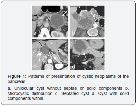
Most of the cystic neoplasms of the pancreas are found incidentally on imaging studies made for another clinical entity, nevertheless the most widely imaging modalities used on the approach of these lesions are the computed tomography, endoscopic ultrasound and magnetic resonance imaging. Imaging features are depicted in Table 1 & Figure 1.


Management
The approach for a patient with a suspicion or finding of pancreatic cystic neoplasms includes a series of studies that will allow to identifying and classifying the type of neoplasm and determining if a surgical resection is feasible. A solid component, main pancreatic duct of more than 10cm in diameter, a cyst with a diameter of more than 3cm, thickened wall or retroperitoneal lymphadenopaties are features that raise the suspicion of a malignancy in this setting. It is also important to analyze (when possible) via EUS the fluid of the cyst to determine cytology and CEA levels which could aid in differentiating a benign and malignant cyst. (Table 2) Studies do not recommend sampling with Endoscopic Retrograde Cholangiopancreatography (ERCP) as a routine procedure [5,6].

Patients with documented pancreatic cyst of less than 3cm. without any solid component or main duct dilation should be followed with magnetic resonance imaging (MRI) in one year and then every two years for a total of 5 years if no significant changes are seen. Patients with cysts of more than 3cm, dilated main pancreatic duct or solid component should be offered for an endoscopic ultrasound guided fine needle aspiration biopsy.
Surgical treatment
Indications for surgical treatment include patients with main duct IPMN and patients with MCN. Surgical techniques include: Total pancreatoduodenectomy with lymph node resection (for patients with high malignant potential) and total or partial pancreatectomy with lymph node sparing for patients without dysplasia or high malignant potential [7]. Valsangkar et al.[2] Studied a series of 851 patients whom underwent surgical resection for pancreatic cyst neoplasms reporting that the most commonly performed procedure was distal pancreatectomy in 44% of patients followed by total pancreatoduodenectomy in 43% and middle pancreatectomy and enucleation in less than 13% [2].
Laparoscopic resection is only recommended for patients with low grade and high grade dysplasia with close follow up every 2 years with MRI for surveillance of remnant pancreatic tissue. Alcohol ablation is only recommended for research protocols and not as an alternative for surgical therapy [5,7].
Conclusion
Surveillance of pancreatic cystic neoplasms is of paramount importance since most of them carry malignant potential. A proper diagnostic approach and early treatment or follow up decreases the chances of progressive malignant disease with the associated dismal prognosis.
For more articles in Open Access Journal of Case Studies please click on: https://juniperpublishers.com/jojcs/
To know more about Open Access Journals Publishers
To read more…Fulltext please click on: https://juniperpublishers.com/jojcs/JOJCS.MS.ID.555594.php


No comments:
Post a Comment