Juniper Publishers- Open Access Journal of Case
Studies
Atypical Presentation of Merkel Cells Carcinoma in Thigh. An Unusual Case
Authored by Andrés Limardo
Abstract
Background: The Merkel Cells Carcinoma (MCC) is a neuroendocrine carcinoma of the skin. MCC within the lymph nodes in the absence of a primary site is rare and has only been reported sporadically
Case report: A male patient of 62 years old consults by right inguinal tumor. Lymphadenectomy is made and it informs carcinoma of cells of Merkel. Hidden primary tumor. After 20 months of follow up appears injury to nodular in internal face of right thigh interpreted like primary tumor. Resection is made. We discuss the disease, the diagnosis and the treatment of these tumors.
Conclusion: The Merkel Cells Carcinoma (MCC) is a neuroendocrine carcinoma of the skin. MCC within the lymph nodes in the absence of a primary site is rare and has only been reported sporadically. Our case may represent a lymph node metastasis from an occult or regressed skin primary, but we cannot preclude the possibility of a primary nodal tumor. It is not known until today as it is “the best” therapeutic choice for these cases
Keywords: Tumor of cells of Merkel; Carcinoma of cells of Merkel; Rare Tumors of soft parts .
Introduction
Merkel Cell Carcinoma (MCC) is a neuroendocrine carcinoma of the skin. Although it is 40 times less common that melanoma malignant has mortality greater than this one, with a 30% in the CCM as opposed to a 15% in melanoma [1]. MCC within the lymph nodes in the absence of a primary site is rare and has only been reported sporadically [2]. A case of tumor of cells of Merkel appears next that it make debut in form hides and after 20 months the diagnosis could be confirmed. We discuss the disease, the diagnosis and the treatment of these tumors.
Case Report
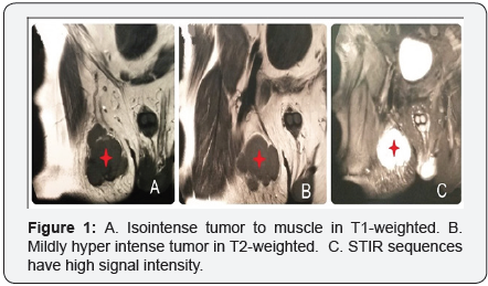
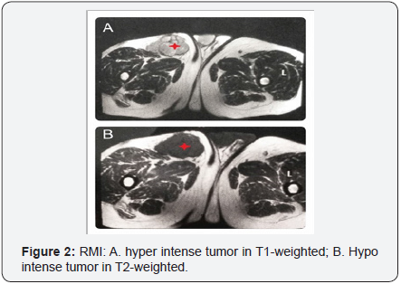
A male patient of 62 years old without relevance antecedents, consult by stony hard tumor in right inguinal region of 2 months of evolution. To the physical examination reveal a mobile and superficial tumor of approximately 35x25mm. It seems inguinal node. The rest of the physical examination in the genital, anal, gluteal zone and inferior tight does not present alterations. Magnetic Resonance Imaging (MRI) of abdomen, pelvis and thighs is realized. It reveals iso intense tumor to muscle in T1-weighted and mildly hyper intense tumor in T2-weightedof 39x28mm in right inguinal region on subcutaneous cellular weave. STIR sequences have high signal intensity. Images around are interpreted like normal nodes (Figure 1). Normal Fibro colonoscopy.

Fine needle aspiration (FNA)
It impresses a neuroectodermic origin with neoplasia of Merkel for which it is necessary Immunohistochemical study to confirm the diagnosis. Surgery is decided. Biopsy by frozen section is made with general anesthesia that informs atipic cells. Inguinal lymphadenectomy is completed.
Histology
Merkel cells carcinoma.
Immunohistochemical study
Cromogranin, sinaptofibrin, CD56 and 20CK 20positive. The injury is interpreted as metastatic carcinoma of cells of Merkel with hidden primary tumor. Reinforcement with 5800cCy is made by Department of Oncology. After 20 months of follow up it appears a violet nodule on skin in internal face of the right thigh that is interpreted like primary tumor of Merkel.
Ultrasound of hi-resolution


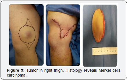
FNA is decided that suspicious tumor informs Merkel cells carcinoma with the antecedents. Surgery is decided. Resection with margins is made, 5cm lateral and 3cm deep and reconstruction with skin local flap (Figure 3). Histology reveals Merkel cells carcinoma confirmed by Immunohistochemical study.
Free margins
After 24 months of follow up the patient does not present relapses.
For more articles in Open Access Journal of Case Studies please click on: https://juniperpublishers.com/jojcs/
To know more about Open Access Journals Publishers
To read more…Fulltext please click on: https://juniperpublishers.com/jojcs/JOJCS.MS.ID.555598.php


No comments:
Post a Comment