Juniper Publishers- Open Access Journal of Case
Studies
Interest of Moringa oleifera in the Treatment of Fungal Mycetoma
Authored by Nomtondo Amina Ouédraogo
Abstract
Fungal mycetoma is inflammatory pseudotumor of subcutaneous soft tissue and bone due to exogenous fungi. it evolution is chronic, often polyfistulated with emission of fungal grains. We report a case of a 44-year-old grower with a fungal mycetoma of the foot with bone damage due to Aspergillus fumigatus. The tumefaction partially regressed after 6 months daily consumption of Moringa oleifera seeds. Also called “miracle tree”, M. oleifera is a shrub, growing in a tropical region, having several properties recognized in traditional medicine, its grains would have an antifungal activity. Doppler, by demonstrating hyper vascularization reflecting the presence of inflammatory remodeling controls the activity of the disease, is an interesting, non-invasive factor for the follow-up of the disease.
Keywords: Eumycetoma; Madura foot; Aspergillus fumigatus; Hypervascularization; Antifungal activity
Introduction
Mycetomas are pathological processes in which agents of fungal or actinomycotic origin emit grains. They are common in tropical and sub-tropical countries. Fungal mycetomas due to fungi and actinomycotic mycetomas due to actinomycetes [1]. Mycetomas are still endemic in the mycetomic belt (on both sides of the fifteenth northern parallel) where Burkina Faso is located and in which the climate makes possible the survival of the agents (long dry season of at least Seven months followed by a short rainy season) [2].
Eumycetomas or fungal mycetomas are inflammatory pseudotumors of subcutaneous soft tissues due to exogenous fungi. They are often polyfistulated with an emission of fungal grains and a chronic evolution. They evolve slowly, determining after certain duration of evolution, osseous attacks that involve the functional prognosis, then visceral attacks sometimes fatal [3]. Treatment poorly codified, recourse to long-term antifungals and surgery in the event of associated bone involvement. We report a case of fungal mycetoma of the foot with bone involvement that partially regressed after 6 month regular consumption of seeds of Moringa oleifera.
Observation
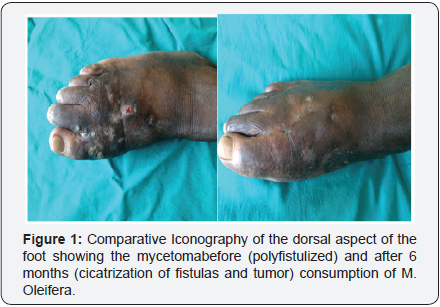
A 44-year-old man Burkinabe, a farmer, consulted the dermatology department of the Raoul Follereau Center for a polyfistulized tumor of the right foot. The anamnesis did not find any notion of local trauma. The disease had begun 5 years ago by a subcutaneous nodule of the sole of the foot, which gradually increased in volume impeding walking. Clinical examination at admission indicated a firm tumefaction of the anterior 2/3 of the foot, going from the back to the sole of the foot about 15cm in diameter, giving rise to black grains of 1 to 2mm in diameter as well as hematic and nauseous (Figure 1). The examination had not found loco-regional and distant nodes. The neurological examination was normal. The rest of the somatic examination was without anomaly. The biological exploration noted only a moderate increase in sedimentation rate to 36mm at the first hour. The hemogram, liver function, renal function, muscle enzymes were normal. Histology compatible with mycetomanoted a polymorphic granulomatous infiltrate (histiocytes, polynuclear neutrophils and lymphocytes) with suppuration foci circumscribing small-sized pigmented grains with irregular contours.


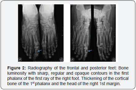
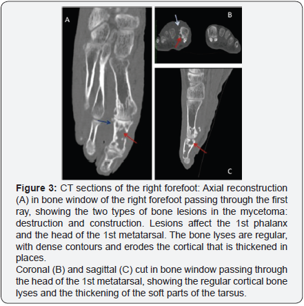
The mycological study concluded a fungal mycetoma by highlighting Aspergillus fumigatus in the tumor collection. The X-ray of the right foot performed in frontal incidence and 3/4 revealed bone luminosity with sharp, regular and opaque contours in the first phalanx of the first radius of the right foot. A thickening of the cortical bone of the 1st phalanx and the head of the right 1st margin was noted. There were no other abnormalities in the bones (Figure 2). CT scan of the feet without and with injection of contrast agent with reconstruction in soft parts and in bone window revealed bone deficits hypodense of cortico-medullary seat, interesting the diaphyso-epiphyseal regions of the first phalange of the right foot and the head of the 1st metatarsal. Their contours were sharp, regular and dense. The sesamoids were dense and atrophic. There was hypertrophy of the soft parts of the forefoot, enhanced after injection of contrast medium (Figure 3). No subcutaneous fluid collection was detected. These imaging aspects led to the conclusion that the 1st metatarsal head of the 1st metatarsal head and the 1st right foot phalanx with the thickening of the bone cortex and thickening of Soft parts of the tarsus facing.

These imaging aspects led to the conclusion that the 1st metatarsal head and the 1st phalanx of the right radius of the right foot were found to have type Ia (Madwell Classification) osteolysis plaques, with thickening of the cortical bone and thickening of Soft parts of the tarsus facing. Subcutaneous hypoechoic formations containing hyperechogenic grains, compatible with a fungic ultrasound. Hyper vascularization at the Doppler may reflect the presence of active inflammatory changes.
A surgical management associated with a terbinafine treatment of 1g per day and antiseptic was proposed but did not meet the consent of the patient, who did not wish to undergo surgery and did not have financial resources for it and no medical insurance to cover him. He interrupted the medical follow-up.
Contacted several times by the healthcare team without success, the patient is finally reviewed after 6 months. The clinical examination noted a significant clinical improvement of the mycetoma lesions with a swelling of the tumefaction compared to the 1/3 middle of the right foot, complete cicatrization of the polyfistulae lesions of the back of the foot. A regression with partial healing of the lesions of the foot is observed (Figure 1 & 4). One notes a persistence of blackish lesions in the middle 1/3 and opposite the plantar face of the toes of the foot. The discomfort with walking has been considerably improved. When questioned, the patient reported initiating a traditional medicine treatment consisting of daily oral intake of 4 seeds of Moringa oleifera (M.oleifera) and applying of the leaves on the lesion for 6 months.

A mycological control (realized for free) of tumefaction was negative as well as the aspergillary serology (realized for free) performed (Ac antiA fumigatus EIA bio rad=0 AU/ml, and Ac anti A fumigates HAI <80).

Doppler ultrasound (realized for free) performed in projection of the plantar face of the right foot, visualized opposite the cutaneous papules, hypoechoic oval beaches. They had different depth levels in the subcutaneous tissue. The deepest ones communicated by pertuis with the more superficial beaches. Within certain deep collections, spontaneously hyperechoic elements with angular contours, measured between 1 and 2mm corresponding to the grains, were visualized. At the Doppler, there is an arterial hyper-vascularization within hypoechogenic rearrangements (Figure 5). An overall thickening of the soft parts of the right forefoot was noted. These subcutaneous hypoechoic formations on the plantar surface of the right foot containing hyperechogenic grains in them, are compatible with a fungic ultrasound. Doppler hypervascularization may reflect the presence of active inflammatory remodeling.
Discussion
The diagnosis of fungal mycetoma of our patient was raised and retained by the anamnestic aspects: long duration of evolution, the notion of trauma not recovered. Indeed, the delay between the occurrence of the disease and the inoculant bite of the germ being very long, the latter is often forgotten [1].
The clinical aspect: the polyfistulized swelling of the foot emitting black grains, the black grains always being of fungal origin [1]. The attack of the foot is usual and defines the classic foot of Madurae.
The histological aspect: a polymorphous granulomatous infiltrate (histiocytes, neutrophilicpolynuclear cells and lymphocytes) with suppuration foci circumscribing small pigmented grains with irregular contours suggestive of mycetoma. This is confirmed by the mycological study which isolated Aspergillus fumigatus confirming the fungal origin.
The first particularity of this case is the isolation of Aspergillus fumigatus from mycology. Indeed, the germs usually incriminated in fungal mycetomas are Madurella Mycetomatis in our context [2,3]. Verma & Kotwal [4,5] studies have reported Aspergillus as the germ involved in eumycetoma [4-7].
The second feature is its clinical regression with M. oleifera seeds consumption, which resulted in complete healing of the lesions of the back of the foot and partial healing of the plantar surface in 6 months.
M. oleifera is a small shrub of the family of Moringaceae, native to India, growing in tropical and subtropical zone, including 13 species, known in traditional oriental medicine for its medicinal properties [8,9]. The species M. oleifera Lam or M. pterogo sperma Gaertn is the most popular in Africa notably in Burkina Faso Called “miracle tree”, all parts of the plant (leaves, bark, seeds, flowers, roots) have significant pharmacological activities: antifungal, hypocholesterolemic, hypoglycemic, antitumor, anti-infectious, antioxidant, antibacterial. Antifungal activity on certain skin fungi such as Trichophyton rubrum, Trichophyton mentagrophytes, Epidermophyton floccosum and Microsporumcanis [10-13]. Thus, in our patient, M. oleifera , by his antifungal properties, would have permitted the complete melting of the tumefaction of the foot, the partial cicatrization of mycetoma of the foot? These questions could serve as a hypothesis for further studies to confirm the antifungal activity of M. oleifera active in the eumycetoma. This anti-fungal activity may prove to be a major step forward in the therapeutic management of fungal mycetomas, especially in developing countries where long-term treatment and the financial inaccessibility of antifungal drugs are a barrier to taking in efficient charge.
The last feature is that the Doppler showed active internal inflammation despite clinical improvement. According to Dikhaye, the use of imaging is essential to the assessment of pretherapeutic extension, but also to the evolutionary follow-up of the bone involvement, essentially and the soft parts of fungal mycetoma, conditioning the indication of medical and surgical treatment as well as the surgical gesture to be realized [14]. The involvement outside the soft parts can be manifested at the articular and extra synovial level as in the case of Thiouri where there was a fusing carpet. Iniesta [15] found the same lesions in the type of destructions and fusion of the leg and foot bones after 20 years of evolution [14,15].
Osteolysis has been described radiologically with varying degrees of developmental outcome. Chronic osteitis (X-ray builder and destructive aspect) and endemic Kaposi disease may be discussed but clinic and mycology were directed towards fungal mycetoma [1,16,17].
At the Doppler, the presence of inflammatory rearrangement always active on the plantar face, indicates the persistence of the activity of the germ in this part of the foot reached. The ultrasound, non-invasive, reproducible, inexpensive appears best adapted in our working context. Mycetomas give characteristic ultrasound images described by Sudanese authors: multiple cavities with thick walls without acoustic reinforcement [18]. It is possible to distinguish, according to these same authors, the action mycetomas of the fungal mycetomas according to the echo graphic aspects. The former have poorly individualized echoes corresponding to small and cement less grain in most species. They are usually grouped together at the bottom of the cavities. Echoes are sharp, shiny, hyper reflective in black fungal fungal mycetomas. Ultrasound makes it possible to delineate the extension of the mycetoma more precisely than the clinical examination [17-19].
Conclusion
This finding reveals a rare fungal mycetoma cause: Aspergillus fumigatus. It also arouses interest in M. oleifera in the management of eumycetoma, which would be appreciable in our working context. Further studies could validate or invalidate this hypothesis. Doppler ultrasound could be an interesting examination for the monitoring and surveillance of fungal mycetoma, which can take several years to manage and help with therapeutic decisions.
For more articles in Open Access Journal of Case Studies please click on: https://juniperpublishers.com/jojcs/
To know more about Open Access Journals Publishers
To read more…Fulltext please click on: https://juniperpublishers.com/jojcs/JOJCS.MS.ID.555600.php


No comments:
Post a Comment