Juniper Publishers- Open Access Journal of Case Studies
Internet Surfing and Multiple Retzius Spaces
Authored by Maulana M Ansari
Abstract
Internet surfing sometimes proves very rewarding with astonishing results. So was the case of chance finding of an old PhD thesis of Mark Hayes published in 1948 (https://deepblue.lib.umich.edu/bitstream/handle/2027.42/49606/1000870105_ftp.pdf?sequence=1). Hayes' thesis largely went unnoticed by the anatomists and surgeons alike, but adds immense value and significance to the author's article published recently (http://www.aimdrjournal.com/pdf/vol3Issue5/SG8_OA_V3N5.pdf ). In addition to the author's report on the laparoscopic anatomy of the Retzius spaces, excerpts of Hayes' thesis warrant general awareness among the anatomists and surgeons, particularly the laparoscopic hernia surgeons doing total extraperitoneal preperitoneal (TEPP) hernioplasty for inguinal hernia. Hayes' thesis for the first time re-evaluated the pelvic anatomy with description of multiple retropubic spaces in the region of Retzius space in a closely graded series of human embryos, fetuses, newborn infants, and adults.
Keywords: Retzius space; Retropubic spaces; Cadaveric anatomy; Laparoscopic anatomy; TEPP hernioplasty
Brief Communication
The author recently published an article titled -'Retzius and Bogros Spaces: A Prospective Laparoscopic Study and Current Perspectives' [1]. Soon after (September 30, 2017), the author incidentally happened to come across during casual internet surfing the PhD (Anatomy) Thesis of Mark Allan Hayes submitted in 1948 to the University of Michigan, Ann Arbor, USA [2]. His thesis was based on an examination of a closely graded series of human embryos, fetuses, newborn infants, and adults carried out over a period of two years in the gross anatomical laboratories of the University of Michigan Medical School. His whole thesis is a marvellous anatomic treatise. However, an illuminating excerpt from the thesis caught the eyes of the author who strongly felt that this awe-inspiring piece of information regarding the Retzius space has a wealth of applied anatomy and will definitely be worth reading by the anatomists and clinical researchers in general and the laparoscopic hernia surgeons in particular, and is reproduced here: "These facts provide material for simplification of the conflicting views concerning the "space of Retzius" (1858). Many observers have given different boundaries for this space (Hyrtl, 1858; Charpy, 1892; Waldeyer, 1899; Rouviere, '24; Hinman, '37; Callander, '39) necessitating re-evaluation of the anatomy concerned. In view of the several fasciae and interfascial clefts present it is no longer possible to consider the "space of Retzius" as a single anatomical, entity. In its place a new concept of multiple fascia1 spaces in this region is suggested. The most ventral of these spaces is the suprapubic space bounded ventrally by the rectus abdominis muscle, dorsally by parietal fascia, caudally by the pubis [2].
"Next in order is the space bounded ventrally by the parietal fascia, dorsally by the umbilical prevesical fascia and for which the term umbilical vesical prefascial space is suggested. This space has dorsal extensions guided by the dorsal limbs of the umbilical prevesical fascia to the region of the pelvic side of the acetabulum. The next space to consider is bounded ventrally by the umbilical prevesical fascia and dorsally by the umbilical vesical fascia, the umbilical vesical interfascial spare. Another more dorsal space is the supravesical space, previously described (p. 138). The most dorsal space is bounded by the umbilical vesical fascia ventrally and the peritoneum dorsally, the umbilical vesical retrofascial space [2]."
Pages 137-138 of Hayes' Thesis [2] read: "The umbilical vesical fascia (Fig. 2) is a specialization of extraperitoneal connective tissue enclosing the bladder and associated structures. It is a triangularly disposed fascia (Fig. 1), the apex of which may reach to the umbilicus and encloses the urachus and both obliterated hypogastric arteries. Caudally the fascia ensheathes the bladder, seminal vesicles, and prostate gland (Fig. 1 and 2). Local condensations of this fascia form the lateral true ligaments of the bladder and the puboprostatic ligaments. At the apex of the bladder, the umbilical vesical fascia can be opened, demonstrating a conical potential space, the base of which is bladder musculature, the supravesical space (fig. 2) [2]."
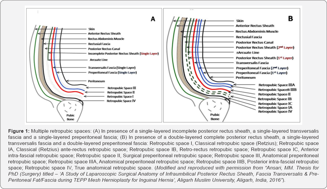
It is not only strange but also unfortunate for the academicians that Dr. Hayes did not touch upon the Bogros space despite ad extenso description of the transversalis fascia, although he referred Bogros in the very first paragraph of the description on the transversalis fascia on page 132 in his thesis [2]. Our observations [1] of the live surgical anatomy during the preperitoneal laparoscopy for inguinal hernioplasty confirmed the cadaveric findings of Dr. Mark Hayes that the Retzius space is not really a single anatomic entity but encompasses a number of potential retropubic spaces namely,
a) True anatomical retropubic space bounded anteriorly by the rectus tendon & pubic bone and posteriorly by the Rectusial fascia,
b) Classical retropubic space bounded anteriorly by the Rectusial fascia & pubic bone and posteriorly by the transversalis fascia, corresponding to the Hayes’ most ventral suprapubic space
c) Surgical preperitoneal retropubic space bounded anteriorly by the transversalis fascia and posteriorly by the preperitoneal fascia, corresponding to the Hayes’ umbilical vesical prefascial space, and
d) Anatomical preperitoneal retropubic space bounded anteriorly by the preperitoneal fascia and posteriorly by the parietal peritoneum, corresponding to the Hayes’ umbilical vesical retrofascial space (Figure 1A).

In presence of the double-layer preperitoneal fascia [3], there was found a potential space between the two layers of the preperitoneal fascia (Figure 1B), corresponding to the Hayes' umbilical vesical interfascial space and the Hayes' supravesical space.
In presence of the complete posterior rectus sheath [4,5], it further divided the classical retropubic space into two potential spaces, or it might mean that the classical retropubic space was bounded posteriorly by both the complete posterior rectus sheath and the transversalis fascia (Figure 1B). Furthermore, in presence of the double-layer complete posterior rectus sheath [5], another potential space was present between its two layers (Figure 1B).
Therefore re-evaluation and re-defining of the retropubic anatomy, especially the Retzius space, is necessitated as originally suggested by Mark Hayes [2], and hence, the laparoscopic TEPP surgeon is strongly advised to be aware of the plethora of these multiple retropubic spaces to avoid a messy frustrating dissection and to be lost into the maze of these multiple retropubic spaces. Presence of these multiple interfascial retropubic spaces explain well the recent observations of Edward Felix that “Initially, the dissection of the extraperitoneal space in the TEP approach tended to be difficult, confusing and therefore hard to learn [6]." I feel obliged to pen down more detailed descriptions of the multiple Retzius spaces with simplified illustrations based on my laparoscopic findings coupled with the cadaveric observations of Mark Hayes in a separate article in near future.
For more articles in Open Access Journal of Case Studies please click on: https://juniperpublishers.com/jojcs/index.php


No comments:
Post a Comment