Juniper Publishers- Open Access Journal of Case Studies
Three Different Benign Breast Lesions of Undetermined Risk in the Same Patient
Authored by Mauricio Camus A
Abstract
We present the case of a 37-years old women with three rare benign lesions of undetermined risk at the same time: a fibromatosis, phyllodes and intraductal papilloma, who was treated by a partial mastectomy. We focus the discussion on fibromatosis, since the other two entities have been previously discussed by our group in other publications.
Keywords: Breast fibromatosis; Phyllodes; Intraductal papilloma
Abbrevations: UOQ: Upper-Outer Quadrant; UQU: Upper Quadrants Union; IQU: Inner Quadrants Union; NSAIDs: Non-Steroidal Anti-Inflammatory Drugs
Introduction
Breast fibromatosis is a rare benign tumor, with uncertain prognosis and management. The objective of this work is to present the case of a patient who had three uncommon benign lesions of the breast and to discuss their management, focusing on the fibromatosis, since the other two entities have been previously discussed by our group in other publications [1,2].
Case Report
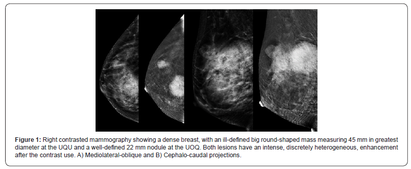
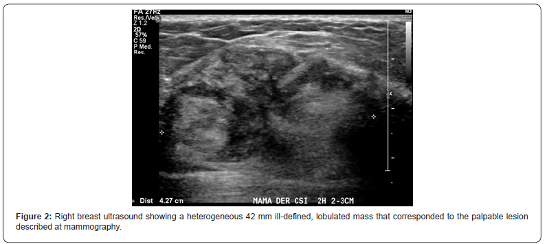
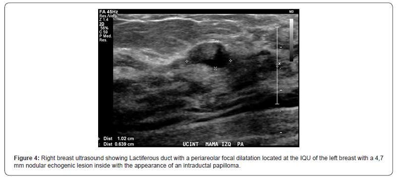
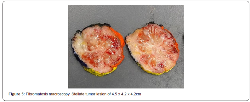
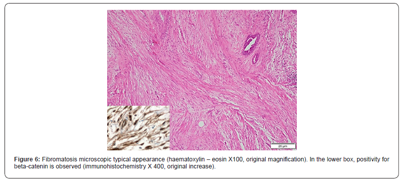
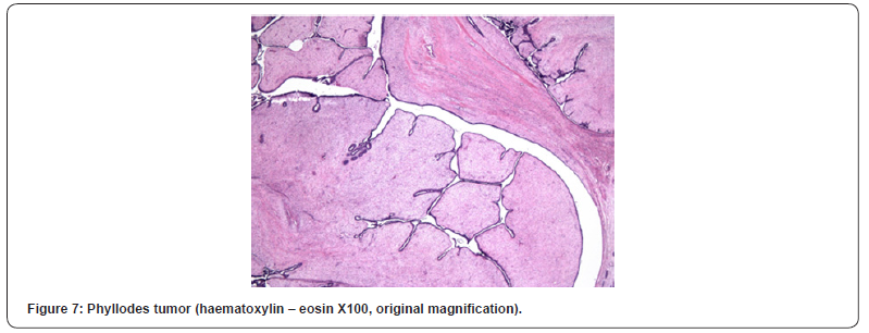
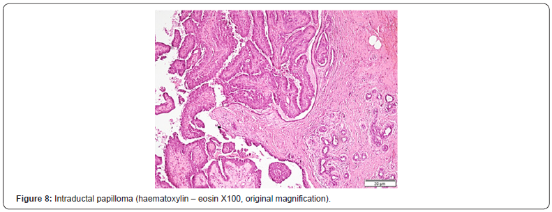
We present the case of a healthy 37 years-old women, with self-detected right breast nodule with 6-months evolution, that was progressively growing and sensitive at touch, associated with erythema and pruritus. She had a familial history of two cousins with breast cancer. At physical exam, there was a hard tumor of approximately 60 x 50 x 30mm at the upper quadrants union (UQU) of the right breast and another 20mm nodule, difficult to separate from the bigger tumor, toward the upper- outer quadrant (UOQ). She was studied with a contrasted mammography that showed a 45mm round-shaped nodule with spiculated edges and early enhancement with contrast, at the UQU (BIRADS-5) and a second 22mm well delimited and moderate enhancing nodule at the upper outer quadrant (UOQ) of the right breast (Figure 1) without invasive-suspected lesions at the left breast. The breast ultrasound demonstrated a 42mm heterogeneous, lobulated mass with imprecise limits and hypoechoic central portion associated with microcalcifications at the UQU of the right breast (Figure 2), a solid oval-shaped well delimited and vascularized 15mm nodule at the UOQ (Figure 3). There was also a 10mm solid-cystic lesion at the inner-quadrants union (IQU) of the left breast (Figure 4). A core biopsy of the two right breast masses was then performed, demonstrating a biphasic fibro-epithelial lesion. The patient underwent a partial bilateral mastectomy of the three lesions, two of them previously marked under ultrasound guidance. The definitive biopsy demonstrated that the lesion of the UQU of the right breast - hour 12, corresponded to a fibromatosis (Figures 5 & 6), and that the other lesion of the UOQ of the right breast was a phyllodes tumor with signs of benignity (Figure 7). In the left breast, an intraductal papilloma was found (Figure 8). The case was discussed at the Oncologic Committee, deciding to follow-up the patient at 6 months with breast ultrasound and contrasted mammography and to offer chronic treatment with celecoxib.

Discussion
Fibromatosis
Fibromatosis is a monoclonal fibroblastic proliferation derived from deep soft tissues. It is characterized by infiltrative growth and local recurrence tendency, without metastasizing capacity, although in 10% of the cases, it could be multifocal [3]. According to the Soft Tissue Tumors classification of the World Health Organization, it corresponds to an intermediate - locally aggressive lesion [4]. Reported incidence is 5-6 cases per million people, corresponding to less than 3% of all soft tissue tumors. It presents mainly in young women, with a peak incidence at 30-40 years, but could appear at any age [5]. Most frequent localizations are the trunk, abdomen and limbs, with the breast being a rare place of appearance, corresponding to 0,2% of all breast tumors. The etiology is unknown, but it has been observed that in 85- 90% of cases there is a mutation at the beta-catenin gene, which related with proliferation and cell survival [6]. Gardner’s syndrome has been described as a risk factor in the context of familial adenomatous polyposis, present in 10-20% of patients, as well as trauma and surgery history. In a retrospective study, up to 25% of the patients had history of breast cancer [7], and it has also been described associated with breast implants [8], which could be explained by a molecular connection between scarring and tissue fibroproliferative alterations [3]. By other side, pregnancy, probably due to exposure to high amounts of estrogen, has been associated with the appearance of this tumor.
Clinically it presents as a firm, painless or slightly painful mass, sometimes with retraction of the skin or nipple, without discharge or palpable axillary adenopathies. In mammography, it is usually confused with breast cancer due to its irregular shape with spiculated margins, just as in ultrasound, where it is seen as a poorly defined hypoechoic lesion with posterior acoustic shadow and an echogenic ring [9]. The diagnosis is confirmed by core biopsy of the lesion.
Unfortunately, fibromatosis has an unpredictable course and there are no prognostic factors that help us determine which treatment is best suited for each patient. The current recommendation as first line treatment is expectant and symptomatic management, especially in asymptomatic patients, with no risky tumors, and those who are unresectable. Observational studies have shown that approximately 50% of the patients may progress in 5 years follow-up [1,10,11] and that 20-30% have spontaneous tumor resolution [12].
Pregnancy is not considered an absolute indication of surgery. Although 40-50% of these tumors progress during this period, it has not been associated with adverse obstetric outcomes. When planning pregnancy in a patient with breast fibromatosis, it is recommended to previously observe the tumor behavior for two years [3,13].
Regarding surgical management, it is recommended to resect the tumor with negative margins, although it has no impact in recurrence [3,14]. Factors associated with recurrence include tumor location (more frequent in the abdominal wall) and the mutation type of the beta-catenin gene [3,15]. It has been described that recurrence 15 months after surgery would be approximately 29% [7]. There are multiple options for medical management. Complete pathological response has been observed with non-steroidal anti-inflammatory drugs (meloxicam, indomethacin and sulindac), however nor the duration of the treatment or the effect are well described [16- 20]. Other option includes using tamoxifen associated or not to NSAIDs. Chemotherapy is reserved for unresectable, fastgrowing or irresponsive to hormonal treatment tumors [21,22]. Radiotherapy can be used as primary management for the tumor, with 70-80% reported long-term local control, as well as an adjuvant treatment, when there are positive margins, the latter being a controversial indication [5,23].
Phyllodes
Phyllodes tumors of the breast are rare fibro-epithelial lesions, corresponding to less than 1% of breast neoplasms. According to the degree of stromal hyperplasia and atypia, the World Health Organization (WHO) categorizes phyllodes tumors into benign, borderline and malignant tumors, however, there is no consensus defining the appropriate surgical margins that ensure a reduction of the risk of recurrence [3].
Intraductal papilloma
Papillary breast lesions are characterized by growing inside the galactophorous ducts and represents a heterogeneous pathology. They are rare, constituting less than 10% of benign breast lesions and less than 1% of malignant neoplasms of the breast. The WHO classification includes 4 entities within the section on intraductal papillary lesions: intraductal papilloma, intraductal papillary carcinoma, encapsulated carcinoma and solid papillary carcinoma [24]. Its management is controversial, although there is consensus that papillomas with atypical characteristics at pathology require surgical excision, because they have a high rate of coexisting carcinoma. There is no agreement regarding the optimal treatment of papillomas without atypia [2].
For more articles in Juniper Publishers | Open Access Journal of Case Studies please click on: https://juniperpublishers.com/jojcs/index.php


No comments:
Post a Comment