Juniper Publishers- Open Access Journal of Case Studies
Optical Illusions Quintuple during Laparoscopic Total Extraperitoneal Preperitoneal (TEPP) TEPP Hernioplasty - Case Report 2
Authored by Maulana M Ansari
Abstract
Anatomical Illusion during non-biliary surgery has not been reported in English literature except for the recent reports by the author [1]. Bilateral laparoscopic total extraperitoneal preperitoneal (TEPP) hernioplasty was performed in a healthy 45-year old man through posterior rectus sheath approach with 3-midline-port technique and direct telescopic dissection in the posterior rectus canal. On right side, five optical Illusions were transiently experienced, viz., rectusial fascia vs. transversalis fascia, primary vs. secondary arcuate line, incomplete vs. complete posterior rectus sheath, lower attenuated part of complete posterior rectus sheath vs. transversalis fascia, superficial layer of double-layered preperitoneal fascia vs. transversalis fascia. Procedure was completed in 2 hours with significant technical difficulties. Simultaneous left TEPP hernioplasty was performed with ease and safety in 45 minutes despite presence of contralateral non-mirror anatomy.
Keywords: Laparoscopic hernioplasty; TEPP/ TEP; Optical Illusion; Anatomical Illusion, Secondary arcuate line; Primary arcuate line; Arcuate line; Posterior rectus sheath; Preperitoneal fascia; Transversalis fascia
Abbrevations: TEPP/ TEP: Total Extraperitoneal Preperitoneal (Hernioplasty); PRS: Posterior Rectus Canal; C-PRS: Complete Posterior Rectus Sheath; IC-PRS: Incomplete Posterior Rectus Sheath
Introduction
Optical Illusion of biliary anatomy is now a well-known human factor for increased incidence of laparoscopic bile duct injury [2]. Anatomical Illusion during non-biliary surgery has not been reported in literature to the best of our knowledge except for a recent report by the author [1,3]. The author experienced a total of five transient optical Illusions during laparoscopic total extraperitoneal preperitoneal (TEPP) hernioplasty in two adult male patients with primary inguinal hernia, and details of one case are presented herein.
Case Report
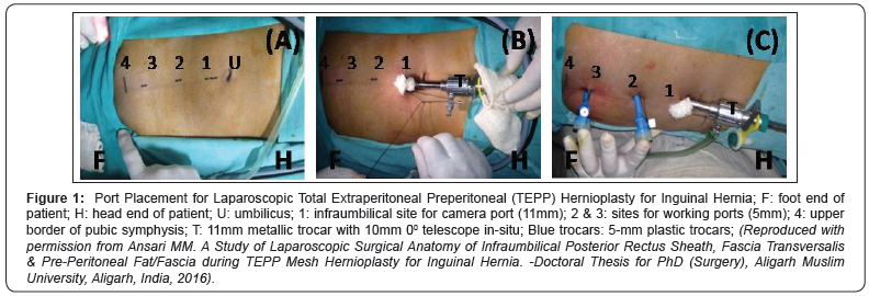
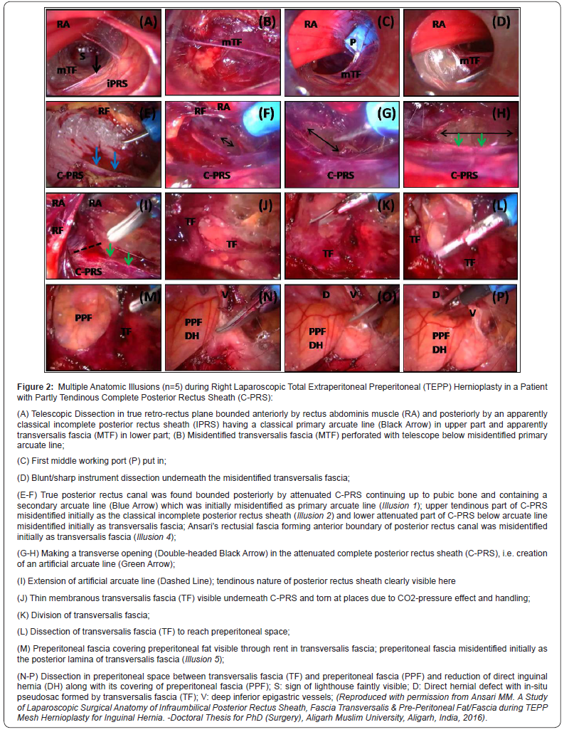
A 45-year-old male labourer (RL #12) with body mass index of 22.6Kg/m2 and ASA grade I presented with bilateral inguinal hernia. Posterior rectus sheath approach was utilized with 3-midline-port technique for bilateral laparoscopic total extraperitoneal preperitoneal (TEPP) hernioplasty. Right side was operated first. Direct telescopic dissection was used to create initial space creation in the posterior rectus canal. On insertion of a 10mm 0º telescope into the posterior rectus canal, the immediate endoscopic view reflected a classical textbook picture as taught in our anatomy classrooms, i.e., the anterior boundary of the posterior rectus canal being formed by the rectus abdominis muscle and the posterior boundary being formed in its upper part by aponeurotic incomplete posterior rectus sheath with a welldefined sharp arcuate line and in its lower part by the transversalis fascia (Figure 1 & 2A).


Further telescopic dissection in this retro-rectus dissection proved rather difficult and bloody due to tearing of tiny vessels supplying both the rectus abdominis muscle and the fascia forming the posterior wall of this plane; the telescopic tip was using to perforate the fascia below the apparent arcuate line, but the plane became a little bloody again (Figure 2B). So, the first working port was then put in, and the blunt/sharp instrument dissection soon rectified the misperception by revealing the presence of a partly tendinous complete posterior rectus sheath extending upto the pubic bone and containing an in-transit secondary arcuate line which was initially misidentified as the primary arcuate line (Illusion 1) (Figure 2C-2D). The upper aponeurotic part of the complete posterior rectus sheath above the secondary arcuate line was misidentified as the classical aponeurotic incomplete posterior rectus sheath (Illusion 2), and the well-defined fascial rectus epimysium called the ‘Rectusial fascia of Ansari’ [4] which was taken down on to the floor of the operating field was initially misidentified as the transversalis fascia (Illusion 3). The lower attenuated part of the complete posterior rectus sheath, i.e., the ‘rectus sheath fascia of Arregui’ [5] was momentarily misidentified as the transversalis fascia (Illusion 4) (Figure 2E). Application of the ‘methods of error reduction’ timely rectified the transient misperception, i.e., the posterior rectus sheath was continuous up to the pubic bone and contained, in addition to a tendinous band called secondary arcuate line, tendinous fibres lower down (Figure 2E-2I) which are never present in the transversalis fascia.
As the primary terminal arcuate line was absent, an artificial arcuate line as described earlier by the author [5-7] for an ergonomic access to the preperitoneal space was surgically created in the complete posterior rectus sheath at the level of the middle port (Figure 2F-2H) when two fascial layers was detected underneath it (Figure 2J-2K). These two fascial layers were initially perceived as the two lamina of a double-layered transversalis fascia in the first glance but further unhurried gentle dissection and application of ‘methods of error reduction’ revealed that the superficial thin membranous fascial layer was really the transversalis fascia proper which formed the pseudosac of the direct inguinal hernia and the deeper diaphanous fascial layer was the preperitoneal fascia/fat (Illusion 5) (Figure 2L- 2M). The superficial thin membranous fascial layer (transversalis fascia) was found readily separable in an avascular fashion from the deeper diaphanous layer (preperitoneal fascia/fat) which covered the direct hernial sac and got easily reduced with it into the abdomen (Figure 2N-2P).
The avascular plane between the transversalis fascia and the preperitoneal fascia, which was earlier reported as the ‘surgical preperitoneal space’ by the author [9,10] was found easily extendable laterally into the ‘surgical preperitoneal space’ of the subinguinal space. These optical Illusions resulted in significant surgical difficulties, leading to completion of the procedure in 2 hours.
During the contralateral TEPP repair on the left side in the same sitting, the anatomical disposition included welldefined rectusial fascia, incomplete posterior rectus sheath of partly tendinous nature, ill-defined primary arcuate line, single diaphanous transversalis fascia and single membranous preperitoneal fascia, and the procedure was completed smoothly and rapidly in 45 minutes.
Discussion
Primary anatomical outcome measures of our doctoral research study have been reported earlier by the author [10-12], re-emphasizing the surgical significance of the anatomic variations of inguinal anatomy sporadically reported in literature since long. Modern newer laparoscopic approaches of surgery offer excellent lighting, high magnification and internal perspective which provide clear visualization of even thinnest fascial layers and tissues not appreciated during gross anatomic dissections because of their hardening and fusion during the process of embalming. However, the laparoscopic environment facilitates occurrence of anatomical Illusions, leading to intraoperative technical difficulties and complications, as documented in relation to the higher incidence of the bile duct injury during laparoscopic cholecystectomy by Way et al. [2] and Stewart & Way [13]. However, the phenomenon of optical Illusion has not been explored in the non-biliary surgery except for a recent report by the author [1]. Highly variable multifascial layers of inguinal anatomy are potentially prone to optical Illusion, as highlighted by the present case. Lack of the knowledge of the older literature of the inguinal anatomy and their frequent variations [10-12] in the early part of our study of the TEPP hernioplasty was possibly responsible for our proneness to the optical Illusion. Laparoscopic hernia surgeon who wishes to start TEPP repair is strongly advised first to go through the anatomic literature beyond the traditional textbooks of surgery and anatomy in order to familiarize him/ herself the frequent variations of abdomino-inguinal anatomy. Optical Illusions during TEPP hernioplasty warrant for due cognizance for their potential occurrence to safeguard against the injudicious dissection/ technical difficulty for seamless execution of the procedure with ease, rapidity and safety.
Non-mirror anatomy on contralateral side has been reported in a high percentage from 37.5% to 62.5% by the author [11,12]. However, experience of laparoscopic TEPP hernioplasty on one side may benefit the operator on the contralateral side although there may not be an exact mirror anatomy on the opposite side as was experienced in the present case.
Conclusion
A total of five optical Illusions were documented during the procedure of laparoscopic TEPP hernioplasty in an adult patient with bilateral primary inguinal hernia. Significant surgical difficulties were experienced with prolongation of the operation time. Due cognizance of the potential optical Illusion during TEPP hernioplasty is strongly recommended in order to pre-empt the injudicious dissection and technical difficulties for hassle-free performance with rapidity and safety.
For more articles in Juniper Publishers | Open Access Journal of Case Studies please click on: https://juniperpublishers.com/jojcs/index.php


No comments:
Post a Comment