Juniper Publishers- Open Access Journal of Case Studies
Giant Lipomas of Anterior and Lateral Cervical Region: Management of a Rare Entity
Authored by Omar Iziki
Abstract
Blood is a vital component of human body which is involved in many functions most importantly supplying oxygen along with essential nutrLipomas are the most common mesenchymal tumours of adulthood, infrequently occur in the head and neck region. Lipomas are typically slow-growing tumours, only a few grow to an exceptionally large size. Surgical excision of a lipoma is the gold standard and the definitive treatment modality. In the present study two rare cases of a giant lipoma of lateral and anterior cervical region are presented.
Keywords: Lipoma; Neck; Anterior; lateral, surgery
Introduction
Lipoma is a slow growing benign tumor composed of adipose tissue, that can be observed all over the body. They are generally small lesions, weighing only a few grams, Lipomas that are greater than 10cm in with or more than 1000g in weight are considered giant tumors. In 13% of case, lipomas are seen in head and neck region, and they occur regularly on the back of the neck. Anterior and lateral localization of lipoma in the neck is rare. We report two rare cases of giant anterior and lateral cervical lipoma, with successful surgical excision, followed by a brief discussion on diagnosis and management of the disorder.
Case 1
A 62-year-old man presented with a complaint of painless swelling on the left side of his neck for a long time, Physical examination revealed a soft, painless, giant and mobile mass beginning from the upper part of the jugular region in his neck (Figure 1). He denied dysphonia, odynophagia and dysphagia. His past medical history was significant for type-two diabetes mellitus and hypertension. The CT scan and MRI showed a large mass media to the left sternocleidomastoid muscle extending from the level of the left mastoid tip to the level of the left clavicle. The lesion extended along the anterior and posterior aspects of the left sternocleidomastoid muscle with a distinct thin capsule and septa. It measured about 08cm (anterior-posterior dimension) x 6.3cm (transverse dimension) x 11m (cephalocaudal dimension). It was well circumscribed without evidence of invasion of adjacent structures. The mass was homogenous and had a signal consistent with fat on all sequences. Fine-needle aspiration biopsy (FNAB) was performed, and the cytopathological diagnosis was lipoma. The patient was operated under general anesthesia. A distinct capsule was identified around the lipoma, which facilitated surgical removal (Figure 2). The lesion was dissected bluntly from the internal jugular and common carotid artery without complications. Final histopathological examination confirms the diagnostic of lipoma.
Case 2
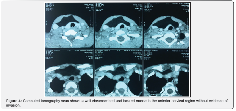
A 11-year-old male, without important pathological antecedents, presented to our ENT clinic, at Casablanca University hospital, with a four -years history of progressive anterior neck swelling (Figure 3), which gradually enlarged in size, without dyspnoea, dysphagia or any complain related to her voice. A physical examination revealed a mobile mass with distinct contours and thickened skin overlying its apex, it was extended from the cervical region to the upper mediastinal region. A CT of the soft tissue of the patient’s neck was recommended. The CT revealed a 14cm × 06cm mass in transverse and longitudinal dimensions, respectively (Figure 4). The mass was well circumscribed and located in the anterior cervical region without evidence of invasion of adjacent structures. The signal intensity of the mass in the CT was homogeneous without internal septations or nodularity.

After general anesthesia was induced, a large mass approximately 15cm in length was excised in its entirety using a minimal incision (Figure 5).
The patient was discharged from the hospital the same day of the surgery. pathological examination revealed a 15cm × 12cm encapsulated tumour. It contained mature adipocytes with no cellular atypia. Numerous fibrous bands through the specimen were evident. The final pathological diagnosis was lipoma.
Discussion
Lipomas are the most frequent mesenchymal tumours of adulthood, it represents approximately 5% of all benign tumours, and they may occur anywhere on the surface of the body. But cervical localization is infrequent [1]. The highest incidence of lipomas occurs between the fifth and sixth decades of life, and lipomas are more common in obese individuals [2]. Although lipomas elsewhere in the body are twice as common in females as in males. In head and neck region, posterior cervical space is the commonest site. We reviewed the literature and saw very few cases of anterior or lateral cervical giant lipoma. Usually they are slow growing and solitary tumors, less than 5cm. Sanchez et al. was defined a giant lipoma as a lesion that exceeding 10cm in one dimension or weighs a minimum of 1000g [3].
Intraoperatively, lipomas may be seen as soft, yellow, shiny, smooth, mobile, encapsulated and occasionally lobulated subcutaneous masses [1]. Microscopically, the lesion is composed of mature adipocytes arranged in lobules, mostly surrounded by a fibrous capsule. Occasionally, the capsule is absent and lipoma infiltrates into muscle, in which case it is considered as an infiltrating lipoma. Rarely malignant transformation of lipoma into liposarcoma has been described. Differentiation of lipoma from liposarcoma may be difficult [2].
Although the etiology of lipomas is not exactly known [4], they are associated with hypercholesterolaemia, trauma, and obesity. Syndromic forms of lipoma have been described, especially in Gardner’s syndrome, Madelung’s disease, and Dercum’s disease. Our 3 cases do not present hypercholesterolaemia, a history of trauma, or obesity [4].
Clinically, vary greatly depending upon the lesion’s size, location and rate of growth. Usually, theses benign tumors are slow growing, painless, mobile, non-fluctuant, soft masses and are generally well encapsulated [1]. Lipomas can be singular or multiple and are typically asymptomatic unless, in the neck region, giant lipoma are primarily a cosmetic problem, they can compress neurovascular or upper airways structures, and may cause dyspnoea and others fuctional symptoms [2].
Radiologic evaluation of lipomas is useful for diagnosis and surgical planning. CT or MRI before excision may provide important information in distinguishing a fatty benign lesion from a liposarcoma before a surgical approach is made [1]. Surgical treatment options of lipomas include excision or liposuction-assisted removal. Excision is more commonly used because of its lower recurrence rate. Liposuctionassisted removal may be advocated to avoid the resulting scar and potential damage to adjacent structures during excision [3]. Complications after excision of a lipoma most commonly include hematoma, followed by seroma, ecchymosis, infection, deformity, injury to adjacent structures, excessive scarring and fat embolus. Lipomas do not have high recurrence rates. Simple lipomas recur 5% locally [2]. Recurrence is related to incomplete excision or infiltrative type of lipoma [4].
Conclusion
In summary, giant lipomas of the head and neck are uncommon. The surgeon should be able to differentiate benign lipomas from liposarcomas. Diagnostic aids include CT scan, MRI, Fine needle aspiration cytology, and open biopsy. Surgical excision is the preferred treatment with low recurrence rates.
To know more about Juniper Publishers please click on: https://juniperpublishers.com/manuscript-guidelines.php
For more articles in Open Access Journal of Case Studies please click
on: https://juniperpublishers.com/jojcs/index.php
Giant Lipomas of Anterior and Lateral Cervical Region: Management of a Rare Entity
Authored by Omar Iziki
Abstract
Blood is a vital component of human body which is involved in many functions most importantly supplying oxygen along with essential nutrLipomas are the most common mesenchymal tumours of adulthood, infrequently occur in the head and neck region. Lipomas are typically slow-growing tumours, only a few grow to an exceptionally large size. Surgical excision of a lipoma is the gold standard and the definitive treatment modality. In the present study two rare cases of a giant lipoma of lateral and anterior cervical region are presented.
Keywords: Lipoma; Neck; Anterior; lateral, surgery
Introduction
Lipoma is a slow growing benign tumor composed of adipose tissue, that can be observed all over the body. They are generally small lesions, weighing only a few grams, Lipomas that are greater than 10cm in with or more than 1000g in weight are considered giant tumors. In 13% of case, lipomas are seen in head and neck region, and they occur regularly on the back of the neck. Anterior and lateral localization of lipoma in the neck is rare. We report two rare cases of giant anterior and lateral cervical lipoma, with successful surgical excision, followed by a brief discussion on diagnosis and management of the disorder.
Case 1
A 62-year-old man presented with a complaint of painless swelling on the left side of his neck for a long time, Physical examination revealed a soft, painless, giant and mobile mass beginning from the upper part of the jugular region in his neck (Figure 1). He denied dysphonia, odynophagia and dysphagia. His past medical history was significant for type-two diabetes mellitus and hypertension. The CT scan and MRI showed a large mass media to the left sternocleidomastoid muscle extending from the level of the left mastoid tip to the level of the left clavicle. The lesion extended along the anterior and posterior aspects of the left sternocleidomastoid muscle with a distinct thin capsule and septa. It measured about 08cm (anterior-posterior dimension) x 6.3cm (transverse dimension) x 11m (cephalocaudal dimension). It was well circumscribed without evidence of invasion of adjacent structures. The mass was homogenous and had a signal consistent with fat on all sequences. Fine-needle aspiration biopsy (FNAB) was performed, and the cytopathological diagnosis was lipoma. The patient was operated under general anesthesia. A distinct capsule was identified around the lipoma, which facilitated surgical removal (Figure 2). The lesion was dissected bluntly from the internal jugular and common carotid artery without complications. Final histopathological examination confirms the diagnostic of lipoma.
Case 2
A 11-year-old male, without important pathological antecedents, presented to our ENT clinic, at Casablanca University hospital, with a four -years history of progressive anterior neck swelling (Figure 3), which gradually enlarged in size, without dyspnoea, dysphagia or any complain related to her voice. A physical examination revealed a mobile mass with distinct contours and thickened skin overlying its apex, it was extended from the cervical region to the upper mediastinal region. A CT of the soft tissue of the patient’s neck was recommended. The CT revealed a 14cm × 06cm mass in transverse and longitudinal dimensions, respectively (Figure 4). The mass was well circumscribed and located in the anterior cervical region without evidence of invasion of adjacent structures. The signal intensity of the mass in the CT was homogeneous without internal septations or nodularity.

After general anesthesia was induced, a large mass approximately 15cm in length was excised in its entirety using a minimal incision (Figure 5).
The patient was discharged from the hospital the same day of the surgery. pathological examination revealed a 15cm × 12cm encapsulated tumour. It contained mature adipocytes with no cellular atypia. Numerous fibrous bands through the specimen were evident. The final pathological diagnosis was lipoma.
Discussion
Lipomas are the most frequent mesenchymal tumours of adulthood, it represents approximately 5% of all benign tumours, and they may occur anywhere on the surface of the body. But cervical localization is infrequent [1]. The highest incidence of lipomas occurs between the fifth and sixth decades of life, and lipomas are more common in obese individuals [2]. Although lipomas elsewhere in the body are twice as common in females as in males. In head and neck region, posterior cervical space is the commonest site. We reviewed the literature and saw very few cases of anterior or lateral cervical giant lipoma. Usually they are slow growing and solitary tumors, less than 5cm. Sanchez et al. was defined a giant lipoma as a lesion that exceeding 10cm in one dimension or weighs a minimum of 1000g [3].
Intraoperatively, lipomas may be seen as soft, yellow, shiny, smooth, mobile, encapsulated and occasionally lobulated subcutaneous masses [1]. Microscopically, the lesion is composed of mature adipocytes arranged in lobules, mostly surrounded by a fibrous capsule. Occasionally, the capsule is absent and lipoma infiltrates into muscle, in which case it is considered as an infiltrating lipoma. Rarely malignant transformation of lipoma into liposarcoma has been described. Differentiation of lipoma from liposarcoma may be difficult [2].
Although the etiology of lipomas is not exactly known [4], they are associated with hypercholesterolaemia, trauma, and obesity. Syndromic forms of lipoma have been described, especially in Gardner’s syndrome, Madelung’s disease, and Dercum’s disease. Our 3 cases do not present hypercholesterolaemia, a history of trauma, or obesity [4].
Clinically, vary greatly depending upon the lesion’s size, location and rate of growth. Usually, theses benign tumors are slow growing, painless, mobile, non-fluctuant, soft masses and are generally well encapsulated [1]. Lipomas can be singular or multiple and are typically asymptomatic unless, in the neck region, giant lipoma are primarily a cosmetic problem, they can compress neurovascular or upper airways structures, and may cause dyspnoea and others fuctional symptoms [2].
Radiologic evaluation of lipomas is useful for diagnosis and surgical planning. CT or MRI before excision may provide important information in distinguishing a fatty benign lesion from a liposarcoma before a surgical approach is made [1]. Surgical treatment options of lipomas include excision or liposuction-assisted removal. Excision is more commonly used because of its lower recurrence rate. Liposuctionassisted removal may be advocated to avoid the resulting scar and potential damage to adjacent structures during excision [3]. Complications after excision of a lipoma most commonly include hematoma, followed by seroma, ecchymosis, infection, deformity, injury to adjacent structures, excessive scarring and fat embolus. Lipomas do not have high recurrence rates. Simple lipomas recur 5% locally [2]. Recurrence is related to incomplete excision or infiltrative type of lipoma [4].
Conclusion
In summary, giant lipomas of the head and neck are uncommon. The surgeon should be able to differentiate benign lipomas from liposarcomas. Diagnostic aids include CT scan, MRI, Fine needle aspiration cytology, and open biopsy. Surgical excision is the preferred treatment with low recurrence rates.
To know more about Juniper Publishers please click on: https://juniperpublishers.com/manuscript-guidelines.php
For more articles in Open Access Journal of Case Studies please click on: https://juniperpublishers.com/jojcs/index.php


No comments:
Post a Comment