Juniper Publishers- Open Access Journal of Case Studies
Five Liters of Hematometra in Postmenpausal Women Case of Advance Stage of Endometrial Cancer
Authored by Alsahabi Jawaher
Abstract
We report the case of a 61-year-old multiparous postmenopausal woman with an unusual manifestation of endometrial adenocarcinoma, as she presented with vague abdominal pain, CT and MRI showed hematometra and ovarian cyst with suspicious of malignancy. Evacuation of 5 liters of uterine contents was done followed by laparotomy and staging. histopathology was endometrial endometrioid adenocarcinoma with metastasis to the ovary.
Keywords: Endometrial adenocarcinoma, Laparotomy, Hematocolpos, Menarche, Hematometra, Pelvic inflammatory disease, Menstruation, Menopause, Pyometra, Surgery
Introduction
The causes of intrauterine fluid collections are age-related. In infancy and at the menarche, hematometra and hematocolpos may occur [1-3]. Between menarche and menopause, uterine fluid collections are usually related to pregnancy or its complications, pelvic inflammatory disease, or menstruation [4]. After menopause, hematometra and pyometra may occur. It was our impression that fluid collections were significantly associated with carcinoma of the uterus; indeed, several of the cases of hematometra and pyometra reported in the literature were associated with uterine cancer [5-8]. There are many reported cases of hematometra in postmenopausal and it was associated with malignancy, few of them reported the etiology was endometrial cancer, most of them had another cause like prior surgery, cervical procedure and radiation.
Case Report
61 years old, P4+3, known hypertensive in atenolol, denied any surgical procedure, postmenopausal for five years never had hormonal replacement, referred to gyne-oncology as increase in her chronic abdominal pain for the last 2weeks, which was vague abdominal and back pain associated with dysuria, no GI symptoms no post-menopausal bleeding or discharges. With ultrasound report of Endometrial thickness of 21mm and query cervical cyst.
On examination
Looks well not in pain, BMI 32.7, Vital signs were normal. Abdomen soft lax pendulous abdomen, difficult to ass masses. Vulva is normal, vagina 1-2cm long only atrophic, cervix not seen, per-rectal examination firm non-tender mass felt in posterior cul de sac.
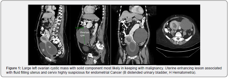
Regarding her investigations; Tumor markers: CA125 was 69.8, CA 19.9 was 50.2, HCG 0.887 CT with oral and rectal contrast was performed, showed Large cystic lesion arising from the left ovary with peripheral solid enhancing component measuring 9 x 14.5 x 15.4cm in AP, transverse and craniocaudal dimensions respectively.
HThe uterus is seen dilated and filled with fluid with small soft tissue enhancing component seen superiorly. The cervix is seen significantly dilated and filled with a fluid measuring 8.1 x 8.6 x 12cm, no soft tissue component seen. There is no local or distant lymphadenopathy seen. No free fluid. No suspicious peritoneal deposits noted. Liver is homogeneously enhancing with no focal lesion. Portal and hepatic veins are patent. Spleen, pancreas, and both adrenals are unremarkable.
Both kidneys demonstrate mild to moderate hydronephrosis and bilateral ureteric dilatation caused by the mass effect of the cervical dilatation. Otherwise both kidneys are unremarkable (Figure 1).
MRI done showed
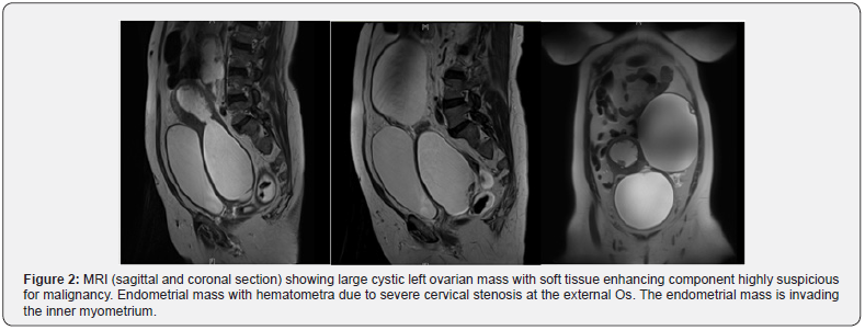
A large left ovarian cystic mass with enhancing solid component measuring 14.2 x 8.7cm at maximum axial plan dimensions. The right ovary appears unremarkable. There is endometrial enhancing soft tissue mass with evidence of invasion to the inner myometrium by less than 50% with no evidence of tumor extension to the cervix.

There is dilatation of the uterus and cervix with bloody fluid and clots with extensive blunting of the cervix extend to the upper vagina probably due to severe stenosis of the external cervical Os. The maximum longitudinal and axial dimension of the endometrial/cervical bloody fluid is about 17.5 x 7.4cm (Figure 2).

Values are expressed as mean ± SD unless otherwise stated. Spearman correlation coefficient (rho) were calculated for non-parametric linear association between different DUS parameters and foot volumetry measurements. p-values < 0.05 were considered significant. Statistical analyses were carried out using SPSS 24.0 for Windows (Armonk, NY: IBM Corp.).
Had a surgery, started by dilating the cervix using fine hegar dilator, then evacuation of intrauterine product around 5 liters of blood drained, in order to shrink the uterine size to have a smaller abdominal incision. Then surgery continued as laparotomy hysterectomy bilateral salpingo-ophorectomy, infracolic omentectomy, left ovarian cyst found ruptured.
Final pathology came as endometrial endometrioid adenocarcinoma FIGO GRADE 1, tumor invade less than 50% of myometrial thickness. left ovary is involved by metastatic carcinoma. no lymph-vascular invasion identified. myometrial adenomyosis and intramural leiomyoma, cervix, fallopian tubes, right ovary and omentum all are unremarkable. Staged as stage 3a (according to FIGO classification), planned to start chemoradiotherapy to be followed by adjuvant chemotherapy.
Discussion
in the postmenopausal patient, fluid in the endometrial cavity has been correlated with gynecological malignancy, but deferential diagnosis and the actual risk of malignancy associated with this finding have not been well defined [4-8].
The case we were encountered she presented with vague symptoms, but clinical examination with help of imaging, diagnosis achieved. In fact, Delay in the diagnosis of uterine tumors usually results from the absence of prominent symptoms, such as abnormal uterine bleeding [9].
Regarding modality of imaging, Transvaginal sonography, allows better visualization of the endometrium in the presence of hematometra and therefore should be used to seek endometrial lesions when hematometra is detected through transabdominal sonography [10]. When adequate vaginal access is impossible because of severe vaginal stenosis, transperineal sonography can be used to visualize the lower genital tract [11]. But in our case ultrasound was confusing, most likely because of huge uterine distention, which made localization for the origin difficult by ultrasound. MRI and CT were modality of choice especially with presence of ovarian mass as well.
Although the degree of association varies among published studies, the presence of uterine fluid collections in postmenopausal women is suggestive of uterine malignancy. Breckenridge et al. [12] found of the 17, 16 (94%) were found to have active carcinoma involving the uterine corpus or cervix. Eleven (65%) had cervical stenosis due to previous surgery, radiation therapy, or carcinoma. However, Carlson et al14 identified only 5 cancers (2 ovarian, 1 tubal, 1 endometrial, and 1 cervical cancer) (25%) among 20 postmenopausal women with intrauterine fluid collections; the remaining women had benign gynecologic conditions [13]. Sonographic findings such as a nodular or thickened endometrium and increasing volumes of fluid seem to correlate with the risk of gynecologic cancer.
McCarthy et al. [14] identified only two cancers (both endometrial) among eight postmenopausal women (25%) with fluid collections; the remaining six had benign endometria. Other reports have attested to the relationship between intrauterine fluid and malignancy, whereas some have cautioned that uterus obstructed by cervical stenosis may fill with fluid and distend from noncancerous etiologies [2,4,15-17].
In the case we presented, CT and MRI were showing large volume of hematometra and suspicious features of malignancy regarding endometrium and ovary. Risk of malignancy appears to correlate with increasing volumes of fluid and other ultrasound - detected alterations, such as nodular or thickened endometrium or clinical symptoms such as discomfort or bleeding [12].
Our case had the maximum uterine contents of reported cases of hematometra, largest volume reported before was 1400ml in Breckenridge et al. [12] 13 case report and it was case of endometrial cancer case.
Conclusion
The sonographic demonstration of a distended uterine cavity in a postmenopausal woman suggests carcinoma involving the uterus. Evaluation by further imaging and obtaining histopathology is mandatory to reach appropriate diagnosis.
To know more about Juniper Publishers please click on: https://juniperpublishers.com/manuscript-guidelines.php
For more articles in Open Access Journal of Case Studies please click on: https://juniperpublishers.com/jojcs/index.php


No comments:
Post a Comment