Juniper Publishers- Open Access Journal of Case Studies
Metachronous Pancreatic Metastases of a Bilateral Renal Cell Carcinoma : A Case Report
Authored by Soufiane Ennaciri
Abstract
We report the case of a 57-year-old patient who developed two pancreatic metastases eight years after surgical treatment of a bilateral renal cell carcinoma. A subtotal pancreatectomy with splenectomy was performed. Three years later, the patient is in good general condition and free from any tumor recurrence. Pancreatic metastases of kidney cancers are rare and onset usually late. They are most often fortuitous discovery during the surveillance of the neoplastic disease. The curative treatment is most often surgical.
Keywords: Pancreatic metastases; Bilateral renal cell carcinoma; Nephrectomy; Pancreatectomy
Introduction
Adult kidney cancer represents 3% of all cancers and the third urological neoplasm [1]. Metastases of renal cancers are usually located in the lung, liver or bones. Secondary pancreatic localization are unusual [2].
Case Report
A 57 years-old man, with a history of smoking, presented to the emergency room for a lower back pain and hematuria with blood clots.
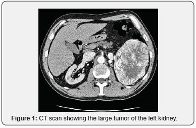
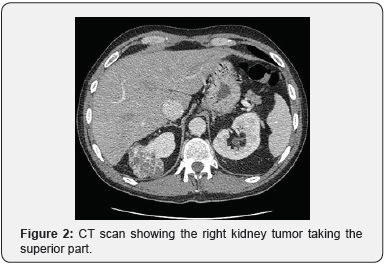
The uroscan by computed tomography showed a bilateral kidney tumor. The left one was very large measuring 12x8x9cm taking up the lower two-thirds of the kidney. There was also a lymphadenopathy and no left renal vein thrombosis. The right kidney was occupied by an heterogenous polar superior tumor, measuring 4.3x5.1x5cm. Screening for metastasis was normal.

The patient under went at first a left radical nephrectomy. The anatomopathological study revealed a clear cell renal cell carcinoma : Fuhrman grade 3, stage pT2N0. Two weeks later, a right partial nephrectomy was performed and histological analysis found a clear cell renal cell carcinoma : Fuhrman grade 2, stage pT1N0 (Figure 1).
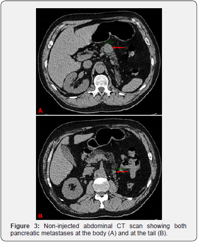
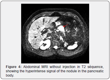
The survey was by CT without injection and MRI as the patient developed renal failure. Two years after nephrectomy, the imaging revealed two suspicious pancreatic nodules, on the body and the tail measuring 3 and 1cm respectively (Figure 2).

Endoscopic ultrasound with biopsy confirmed the metastatic nature of the pancreatic lesions. So the treatment was as à subtotal pancreatectomy with splenectomy. Besides the appearance of diabetes, there was no tumoral recurrence three years after the surgery.
Discussion

Clear cell renal cell carcinoma represents up to 90% of adult kidney cancers. Risk factors include mainly smoking and obesity [3]. The tumor is usually unilateral. In case of bilateral localization, a hereditary disease should be suspected and the surgical approach should be conservative [4] (Figure 3-5).

Pancreatic metastases of renal carcinoma are rare, representing 2 to 5% of pancreatic malignancies [5]. The average time between the nephrectomy and the metastasis diagnosis is 8 years [5] as it was the case for our patient.
Pancreatic metastasis are often asymptomatic. However, they can cause abdominal pain, jaundice, gastrointestinal bleeding, weight loss, exocrin or endocrin pancreatic insuffiency and even acute pancreatitis [5,6].
In most cases, there are discovered incidentally in imaging. Indded, doppler ultrasound and abdominal CT are firstline examination, they allow us to see the heterogenous and hypervascular character of the pancreatic lesion. RMI is of great help in case of renal failure. The result are similar to the CT scan but with better detection of the lesion before injection of contrast product wish is hypoitense on T1 and hyperintense on T2-weighted images [7].
The diagnosis can only be confirmed with the anatomopathological examination [8]. Which can be done after a guided CT, endoscopic ultrasound biopsy or after surgical excision [7].
Surgical excision is currently the best curative treatment. Depending on the lesions topography, a pancreaticoduodenectomy or a spleno-pancreatectomy can be performed [9,10].
Conclusion
Pancreatic metastases of clear cell renal cell carcinoma are rare and can occur many years after the nephrectomy, hence the interest of a careful surveillance. The accessebility of medical imaging leads to the early diagnosis at an asymptomatic stage, thus making them accessible for a surgical treatement, allowing a good survival rate in the long.
To know more about Juniper Publishers please click on: https://juniperpublishers.com/manuscript-guidelines.php
For more articles in Open Access Journal of Case Studies please click on: https://juniperpublishers.com/jojcs/index.php


No comments:
Post a Comment