Juniper Publishers-Open Access Journal of Case Studies
Wolf`s Isotopic Response: A Case of Metastatic Gastric Adenocarcinoma
Authored by Emadodin Darchini-Maragheh
Abstract
Wolf’s isotopic response is defined as the occurrence of a new skin disease at the site of a previous treated or untreated skin disease, mostly herpes zoster. A 70-year-old man, presented with persistent lesions at the same location of previously healed herpes zoster infection. Histopathology revealed diffuse infiltration of signet-ring form neoplastic cells. Further evaluations confirmed gastric adenocarcinoma in the patient. Rare etiologies of Wolf`s isotopic response should be known by physicians and early histopathologic assay when face to this phenomenon in order to early diagnosis is essential.
Keywords: Wolf`s isotopic response; Herpetic disease; Hematologic malignancies; Gastric fundus ulcer; Perigastric lymph nodes; Thrombophlebitis, Mucinous adenocarcinoma
Case Report
Wolf`s Isotopic Response (WIR) refers to a new skin disease, appears at the same location (isotopic) of previously healed and unrelated skin condition. The initial skin disease in most of cases is herpes zoster [1]. WIR was first described in 1955. There is fine difference between WIR and Koebner’s isomorphic response. Koebner’s isomorphic response describes the appearance of same existing skin lesion arising at a site of damaged or traumatized skin [2]. Whenever the isotopic response follows a herpetic disease, the phenomenon is named as Post-Herpetic Isotopic Response (PHIR) [2].
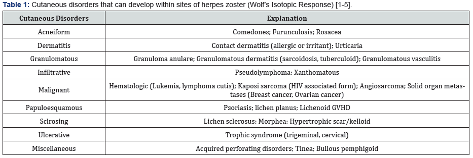
After the initial skin herpes disease has healed, a wide variety of dermatoses can be detected on the same site, which mainly include granuloma annulare, furunculosis, pseudolymphoma, sarcoidal granuloma, tuberculoid granuloma, granulomatous vasculitis, skin malignancies (non-melanoma and melanoma skin cancers), hematologic malignancies (leukemia, lymphoma), solid organ malignancies, etc. [1-4]. Table 1 demonstrates cutaneous disorders that can develop within sites of herpes zoster. Skin metastasis has been infrequently reported to present as WIR.

Case Report
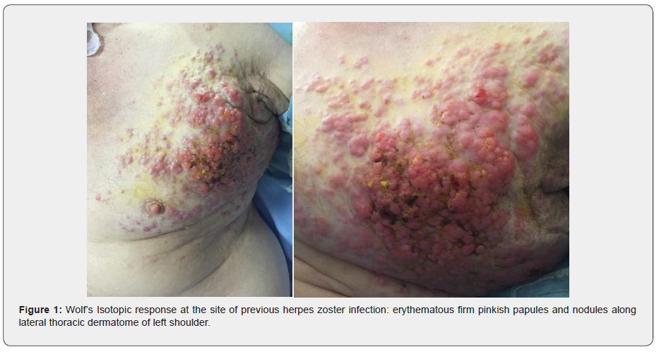
A 70-year-old man was admitted at the department of Internal Diseases due to severe lethargy and weight loss and examined by a dermatologist through a consultation. The patient presented with a two-month history of erythematous firm pinkish papules and nodules along lateral thoracic dermatome of his left shoulder, in the same location as previously healed herpes zoster infection, which was resolved after treatment with oral valacyclovir one year ago (Figure 1). No vesicle or blister was observed. No pain or dysesthesia was present. Further skin examination revealed no related dermatosis. The patient mentioned history of neglected gastric fundus ulcer since 2 years ago.


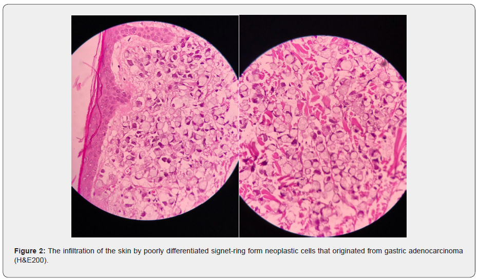
Regarding physical examination, two incisional skin biopsies were performed, suspicious to skin metastasis or granulomatous lesions. Histological assays revealed diffuse infiltration of signetring form neoplastic cells within the superficial and deep dermis as well as excess extracellular mucin formation and lymphovascular embolic foci, reminiscent of mucinous adenocarcinoma (Figure 2). The patient underwent lower and upper endoscopy in the same admission and histologic examination of resected gastric fundus mass revealed poorly differentiated adenocarcinoma with signet-ring cells.
The tumor had invasion into the serosa and perigastric lymph nodes of both curvatures of the stomach, defined as metastatic carcinoma. The patient was classified as stage IIIA (T3N2M0) and referred to the oncologist for appropriate treatment. The skin lesion was shrunk after six sessions of adopting chemotherapy, nevertheless, the patient succumbed to the disease one year after diagnosis of the carcinoma. Written informed consent had been obtained prior to the death.
Discussion
In most cases of WIRs, the initial dermatosis is herpes zoster, known as PHIR, although other initial dermatosis may be varicella, herpes simplex, or thrombophlebitis [5]. Metastasis of internal organ tumors can be rarely manifests as WIR. Hence, awareness and recognition of this entity, are important for diagnosis.
Among solid organ tumors, breast carcinoma and ovarian carcinoma have been formerly reported to have cutaneous manifestation as PHIR. Previously, Wyburn-Mason has reported cases of breast and laryngeal carcinomas developing at the site of previously healed herpes zoster [6]. More recently, Jaka-Moreno [5] has performed a retrospective study on 9 patients with underlined malignancy who presented by PHIR. Five patients had underline B-cell chronic lymphocytic leukemia, non-Hodgkin lymphoma was diagnosed in two patients, and one had ovarian carcinoma [5].
The pathogenesis of WIR is unknown. Herpes zoster may subsequently damage nerve fibers in the dermis, alter immunity and lead to hyper-reactivity to the inflammatory processes such as granulomatous reaction, or local immunosuppression, leading to tumor infiltration, such as leukemia and other malignancies. Moreover, abnormal angiogenesis caused by the nerve damage may also play role in the pathogenesis [7].
Conclusion
Clinicians should be well informed in all feature of herpes zoster, including PHIR; so, in this literature, we highlight the importance of skin punch biopsies when face with WIR phenomenon. In aggregate, this article especially points out rare malignant etiology of PHIR which can be new and useful. To our knowledge, this is the first reported case of gastric adenocarcinoma manifested as PHIR.
To know more about Juniper Publishers please click on: https://juniperpublishers.com/manuscript-guidelines.php
For more articles in Open Access Journal of Case Studies please click on: https://juniperpublishers.com/jojcs/index.php


No comments:
Post a Comment