Juniper
Publishers- Open Access Journal of Case Studies
Breast Hamartoma: Mammography and Magnetic Resonance Imaging
Authored by Biren A Shah
Abstract
Hamartoma of the breast is an uncommon benign mass of the breast. This painless, mobile breast mass is typically diagnosed using mammography, with the pathognomonic finding of a “breast within a breast.” Additional imagings techniques are used when inconclusive results are found with mammography. This report describes a patient case of a classic hamartoma seen on a screening mammogram and on a breast MRI study.
Introduction
A breast hamartoma is a relatively uncommon, benign, slow growing mass found in women. The incidence of hamartoma of the breast is reported at 0.1-0.7%, but this is likely under diagnosed [1]. Current estimates report an incidence of 1.2% with increased use of mammography [2].
Pathologically, the hamartoma is a well circumscribed benign mass composed of fat, dense fibrous tissue, and glandular tissue. Clinically, they typically present as round, painless, and mobile breasts lumps [3]. Breast hamartomas are generally found in women older than 35 years of age, with size ranging from 1cm to 17cm and are presumed to be due to dysgenesis [4]. Most lesions are asymptomatic, and are typically discovered as incidental findings on screening mammography. If a palpable mass is discovered on physical exam, mammography is the preferred method for diagnosis.
Case Presentation
43 year-old female for a bilateral screening mammogram.


Discussion
Hamartoma of the breast was first described by Arrigoni et al. [5] as breast lesions that were well circumscribed and clinically resembled fibroadenoma. Histologically, these lesions showed glandular elements with preserved ducts, lobules, fibrous stroma, and adipose tissue in variable proportions [5]. In 1978, Hessler et al. [6] described the mammography findings of breast hamartoma as a “breast within a breast” because the hamartoma is completely separate from the rest of the breast. It does not replace breast tissue, but instead pushes the inherent breast parenchyma aside [6].
Diagnosis of breast hamartoma typically occurs with mammography, whether during typical screening as an incidental finding, or when indicated by detection of a palpable mass on physical exam.
On mammography, a circumscribed oval or round mass consisting of fat and fibroglandular tissue is highly suggestive of a hamartoma. It has been described as having a “breast within a breast” appearance with lobulated densities comparable to a “slice of salami.” Other findings on mammography may include a thin, radiopaque pseudocapsule and benign calcifications.
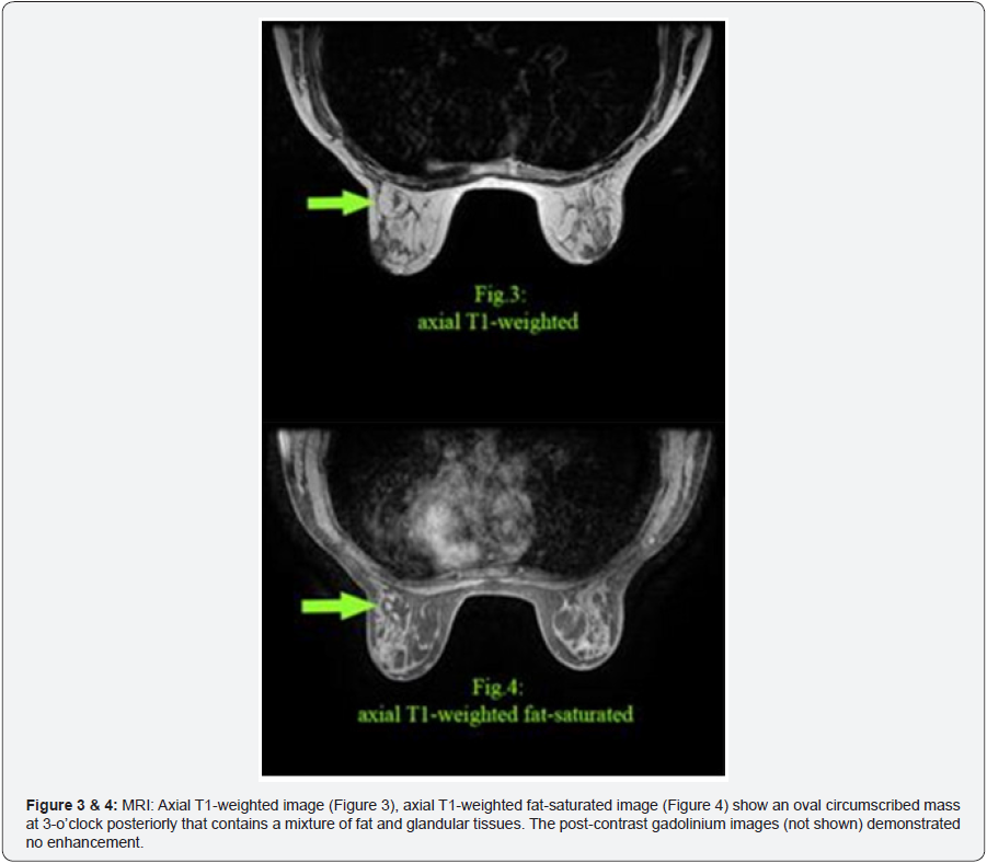
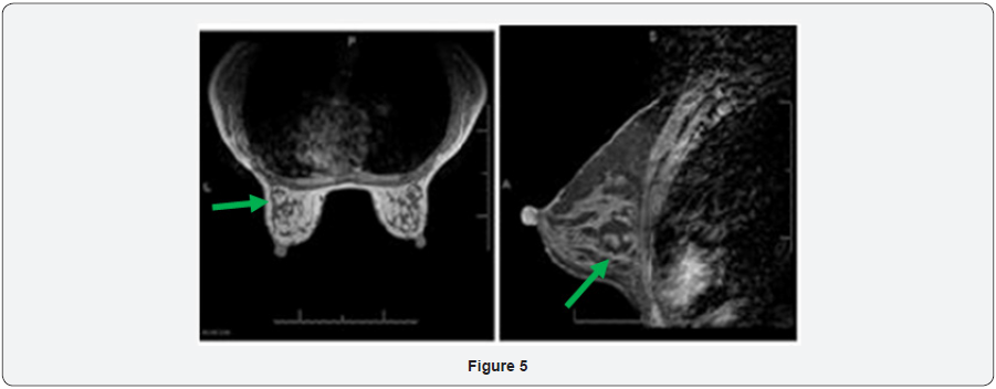
If the typical mammographic signs are not seen and results are inconclusive, additional diagnostic techniques are recommended. If a lesion contains little fat and demonstrates a dominance of fibrous or fibroglandular elements, fibroadenoma or a well circumscribed carcinoma must be ruled out [7]. Additional imaging that can help in diagnosis of breast hamartoma includes ultrasonography, computed tomography, and core biopsy [8]. Magnetic resonance imaging is not commonly used to diagnose hamartoma of the breast but does have characteristic findings. On MRI, hamartomas are well encapsulated with a dark smooth thin rim, oval or round in shape with internal fat densities and heterogeneity. They do not demonstrate significant gadolinium enhancement. MRI findings can provide diagnostic information in a lactating or pregnant woman in whom mammography is not preferred. Erdemet al. [9] described the MRI appearance of a hamartoma as that of an encapsulated mass with fat and fibroglandular signal intensity. The fibro-glandular elements may exhibit some contrast enhancement. Although these other forms of imaging can be utilized, they are not recommended when the mammography findings demonstrate the typical “breast within a breast,” and can even be counterproductive [10].
The characteristic mammographic appearance of a breast hamartoma is pathognomonic and virtually diagnostic. Typically no further follow-up or intervention is required. Excision can be performed if the lesion is symptomatic or if the patient is bothered by the mass. Malignancy with a breast hamartoma is extremely rare and thus aggressive management is not indicated.
For more articles in Open Access Journal of Case Studies please click on: https://juniperpublishers.com/jojcs/index.php


No comments:
Post a Comment