Juniper Publishers- Open Access Journal of Case Studies
Cardiac Arrest in a 45-Year-Old Woman with right Atrial Rhabdomyoma: A Case Report
Authored by Clément Schneider
Abstract
Background: Cardiac rhabdomyoma is very rare in adults, although it is the most frequent cardiac tumor in children.
Case summary: A 45-year-old woman had a cardiac arrest during sclerotherapy for varicose veins. After successful cardiac resuscitation, the patient recovered without neurological damage. Etiological assessment led to the diagnosis of a right atrial cardiac tumor. Surgical excision was undertaken through a minimally invasive thoracotomy on a beating heart with cardiopulmonary bypass. Histological diagnosis was rhabdomyoma.
Discussion: Right atrial rhabdomyoma is exceedingly rare in adults without tuberous sclerosis. Surgical resection can be undertaken to confirm the diagnosis and to prevent potential risk of malignant arrhythmia.
Keywords:Malignant arrhythmia; Right atrial rhabdomyoma; Cardiac tumor; Cardiopulmonary bypass; Uterine adenomyosis; Cardio-circulatory arrest; Transesophageal echocardiogram
Introduction
Cardiac rhabdomyomas are rarely described in adults. Although benign, they can cause arrhythmia or grow to a significant, potentially obstructive, size.
Case Presentation
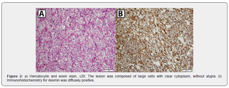
A 45-year-old woman with history of uterine adenomyosis, sural venous thrombosis treated with low-weight molecular heparin five years ago and sclerosis of varicose veins three years ago, had previously been healthy, without any symptoms prior to presentation. Sclerotherapy was performed for varicose veins. Five minutes after the injection of 1.5ml of 0.5% polidocanol in the varicose veins, she presented a sudden loss of consciousness with a cardio-circulatory arrest. External chest compressions were immediately started. Within less than 10 minutes chest compressions, she recovered spontaneous breathing and femoral pulse. On arrival of the emergency ambulance she was unconscious with signs of pulmonary edema. She was therefore intubated, referred to the intensive care unit and placed on noradrenalin 0.9ug/kg/min for initial low blood pressure in the context of sedation, post-cardiac arrest and suspicion of allergic reaction. The initial ECG showed sinus rhythm with tachycardia, large P waves and diffuse flattening of ST segment. She was extubated 24 hours later, weaned of vasoconstrictors after 3 days and recovered without neurological lesion. Cerebral computed tomography and magnetic resonance imaging were normal. Lung perfusion and ventilation scintigraphy showed no evidence of pulmonary embolism. Allergic reaction to polidocanol was ruled out with a negative histamine test. Initial left ventricular function was reduced secondarily to chest compressions and cardiac arrest, but was normal after four days, without left ventricular dilatation or hypertrophy. A right atrial tumor was discovered, attached to the atrial septum without impact on neither right ventricular nor tricuspid function. A transesophageal echocardiogram confirmed the presence of this mass at the foot of the upper vena cava, with a central lower echogenicity, measuring 25x16mm (Figure 1a). Cardiac magnetic resonance imaging confirmed these data (Figures 1b & 1c). No calcifications were noted. T1-weighted magnetic resonance imaging showed a homogenous iso-intense signal compared with surrounding myocardium. Cardiac catheterization was performed. Coronary anatomy was normal, without atherosclerosis, and no tumor blush. Forty-five days after the initial presentation, excision of the tumor was undertaken through video-assisted minimally invasive right lateral thoracotomy and right atriotomy on beating heart with cardio-pulmonary bypass. The mass was attached to the anterior wall of the right atrium near the stoma of the superior vena cava. Macroscopically the tumor measured 4 centimeters in its maximal diameter. It was brownish, moderately heterogeneous but round, smooth and visibly covered with endocardium (Figure 1d). Histological examination (Figure 2) revealed a welldemarcated tumor, consisting of oval or polygonal tumor cells with clear cytoplasm and small nucleus without atypia. Neither tumor necrosis nor mitoses were identified. Some cells had a spider appearance, characterized by perinuclear cytoplasmic radial extensions. PAS staining was positive, showing an abundant intracytoplasmic granulated glycogen. Immunohistochemistry revealed that the tumor cells were positive for desmin (NCLDE- R-11 clone, Ventana, Roche Tissue Diagnostics) and HHF 35 muscle actin (HHF35clone, Dako). The Ki67(Mib1clone, Dako) proliferation index was lower than 1%. All these features lead to the diagnosis of rhabdomyoma. She had neither skin lesions nor familial history suggestive of tuberous sclerosis. Therefore, no genetic testing was performed. Postoperative course was marked by a right pneumothorax regressive with pleural drainage. She was discharged 11 days postoperatively. Four/span> years later she was asymptomatic with normal echocardiogram.

Discussion
Cardiac rhabdomyoma is the most common cardiac tumor in infancy and childhood with a strong association with tuberous sclerosis [1]. These congenital hamartomas are extremely rare in adults [1,2]. Right atrial cardiac rhabdomyomas in adults with no tuberous sclerosis are extremely rare [1,3]. This benign neoplasm showing skeletal-muscle differentiation may be asymptomatic or cause arrhythmias, sudden death [4], ventricular outflow obstruction or even be spontaneously regressive [5]. Left ventricular tumors can cause ventricular arrhythmias and death. In our case, the location of the tumor was peculiar. The probability that the tumor may have temporarily blocked the superior venous return is unlikely. Also, in that specific case, toxicity of polidocanol cannot be completely excluded. Indeed, animal studies have shown that polidocanol injection could cause severe pulmonary edema [6]. Moreover, several cases of polidocanol cardiac toxicity leading to cardiac arrest minutes after foam sclerotherapy have been reported [7,8]. In that case, the discovery of the tumor in our patient could be an epiphenomenon. Some have described spontaneous regression of similar tumors [5] and therefore advocated surgical resection for symptomatic patients only. But, even though cardiac magnetic resonance imaging is peculiarly suited for cardiac tumors assessment [5], it cannot provide the definitive histological diagnosis. Therefore, minimally invasive surgical approach appears an appropriate strategy for diagnosis and treatment of such a lesion [9]. Indeed, histological differential diagnosis from malignant rhabdomyosarcoma is necessary given its significant impact on prognosis and long-term outcomes. Rhabdomyomas are benign tumors but long-term risk of recurrence after resection in adults is unknown.
Conclusion
Right atrial cardiac rhabdomyoma is extremely rare in adults. It can be asymptomatic or cause fatal arrhythmias. Surgical resection can safely be undertaken with minimally invasive techniques.
To know more about Juniper Publishers please click on: https://juniperpublishers.com/manuscript-guidelines.php
For more articles in Open Access Journal of Case Studies please click on: https://juniperpublishers.com/jojcs/index.php


No comments:
Post a Comment