Juniper Publishers- Open Access Journal of Case Studies
The Fibular “Fleck Sign” as Diagnosis of Peroneus Tendons Instability
Authored by Iván Pipa Muñiz
Abstract
Peroneal tendons run along the posterior face of the fibula and its principal stabilizer is the superior peroneal retinaculum (SPR), since they run into. Peroneal tendon subluxation is a rare disorder that is frequently misdiagnosed, and it is caused by the SPR avulsion. It affects mainly young people during high demand sports practice. The injury of the SPR requires surgical treatment because of the undergoing consequences will be instability of peroneal tendons. We present the case of a patient who had a fibular bone avulsion of the SPR (“fleck sign”) while skiing.
Keywords:Peroneal tendons; Superior peroneal retinaculum; Peroneal tendons dislocation; Fleck sign
Introduction
Superior peroneal retinaculum (SPR) is the main stabilizer of the peroneal tendons in the distal third of the fibula. Lesion of the SPR is a cause of peroneal tendons dislocation and it usually requires surgical treatment [1,2]. Eckert and Davi [3] classified the SPR into four types:
a) Type 1 there is an avulsion of the fibular insertion of the SPR.
b) Type 2 there is fibular fibrocartilaginous impeller separation.
c) Type 3 there is a bone avulsion of the fibular SPR insertion.
d) Type 4 there is an avulsion of the calcaneus SPR insertion.
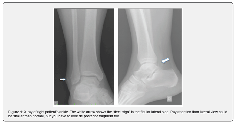
In Type 3, usual X-ray study can show a bone block of the fibular insertion of the SPR known us “Fleck sign”, which is pathognomonic of SPR lesion, which translate an anterior avulsion of the SPR attachment.
In case of this be neglected or ignored, this injury could be misdiagnosed and could not receive adequate treatment, with a dangerous future prognosis, around pain and peroneus tendon’s instability [4,5].Case Report
We present a case report of a 45-year-old male patient with acute right ankle pain after a skiing trauma. He refers a fall without proper boot fixing, and obviously with little inside it movement. As he felt no pain, he finished his practice of the day without find out, and when he took off his boots, he felt that his foot hurt, he could not stand on it and not weight bearing was possible.
Physical examination unveiled swelling and haematoma on the lateral malleolus. Ankle range of motion was limited because of the pain, and neurovascular checking was intact. X-ray made in the emergency room (Figure 1) showed the named “fleck sign” as we showed above.

The fleck sign in the ankle refers to a cortical avulsion fracture of the lateral malleolus. When this appears, the superior peroneal retinaculum is injured, meaning a bone avulsion.
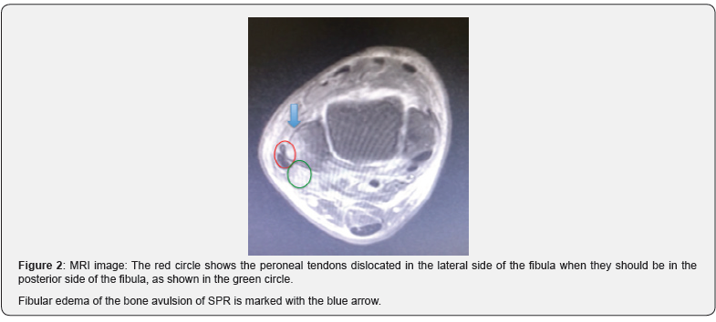
Despite this patognomonical sign we decided to complete the study with an MRI study. The MRI clearly demonstrated the bone avulsion of the anterior peroneal retinaculum, with a lateral peroneal dislocation as well as a longitudinal intrasubstance peroneus brevis tear (Figure 2).

We advised the patient to take surgical intervention and after a correct preop study and signed IC surgery was made. Lateral approach over posterior aspect of the malleolus peroneus was made, over the bone crest. A small bony fragment was found and so, confirmed the avulsion of the SPR. Both tendons, brevis and major, were found anteriorly dislocated through the avulsion. SRP was intact (Figure 3). After an intratendinosus suture of the peroneus brevis by mean of usual stiches, and after that we reduced the peroneus tendons from their anteriorly dislocation (Figure 4) into de retromaleolar groove and reattached the SRP with two suture osseous anchors (Figure 5).
Post-operative treatment was made with a 4 weeks posterior cast and not-weight bearing. After that, 2 more weeks of not weight bearing and passive movement for improvement range of motion was ruled. After 6 weeks, patient started with progressive weight bearing and active motion, working with a specific physiotherapy protocol-based on increasing the weight bearing and movement first of all in swimming pool and progressive till return to play.
Discussion
After ankle trauma, we usually suspect the possible lesion of periarticular ligaments or the presence of a tibial or fibular malleolus bone fracture. We recommend should bear in mind that other structures -such as peroneal tendons or the SPR- might also be affected, if usually it is not very common, a full exploration of these structures must be carried out and, eventually, in case of find it suspect a peroneal tendon dislocation, and surgical treatment will be necessary [6].
In this specific case of SPR, four types of lesions can be identified; in Type 3 of SPR lesions, in X-ray we can see a bone block of the fibular insertion of the SPR which is pathognomonic of the affectation of SPR [1,4].
Alongside the lesion of the SPR, we are likely to find out a lateral or anterior luxation of the peroneal tendons and usually any tear of these tendons (frequently, peroneous brevis) [7]. All together will indicate a need for surgical treatment that in this particular case will consist in suturing and reducing for closing the peroneal Groove of peroneal tendons by mean of a posterior reattachment of the bone block to its origin in the fibula, in order to make or reproduce a bumping post to avoid future peroneus anteriorly displacement. A conscious and selective rehabilitation protocol must be followed [8].
Fleck sign is a radiographic finding in many different body locations. There are similar cases reported in hand [9] (Stener´s Lesion), hip [10] (posterior labral tear), knee (Segond´s Fracture), foot (Lisfranc injuries). In ankle, this fleck sign is usually associated with a medial malleolus injury. For our knowledge there is not any case like this one without any different injuries in middle ankle [1] or talus [4].
Conclusion
The “fleck sign” in the ankle is pathognomonic of bone avulsion of the SPR. It is an uncommon injury that is usually misdiagnosed. Special attention must be paid for this “fleck sign” in the X-ray images because its presence suggests a high probability that peroneal tendons will have been luxated and, consequently, surgical treatment will be needed.
To know more about Juniper Publishers please click on: https://juniperpublishers.com/manuscript-guidelines.php
For more articles in Open Access Journal of Case Studies please click on: https://juniperpublishers.com/jojcs/index.php


No comments:
Post a Comment